

Alimentary System
Stomach, Forestomach - Ulcer
Narrative
Bertram TA, Markovits JE, Juliana MM. 1996. Non-proliferative lesions of the alimentary canal in rats GI-1. In Guides for Toxicologic Pathology. STP/ARP/AFIP, Washington, DC, 1-16.
Full Text: https://www.toxpath.org/docs/SSNDC/GINonproliferativeRat.pdfBetton GR. 1998. The digestive system I: The gastrointestinal tract and exocrine pancreas. In: Target Organ Pathology (Turton J, Hooson J, eds). Taylor and Francis, London, 29-60.
Brown HR, Hardisty JF. 1990. Oral cavity, esophagus and stomach. In: Pathology of the Fischer Rat (Boorman GA, Montgomery CA, MacKenzie WF, eds). Academic Press, San Diego, CA, 9-30.
Abstract: https://www.ncbi.nlm.nih.gov/nlmcatalog/9002563Leininger JR, Jokinen MP, Dangler CA, Whiteley LO. 1999. Oral cavity, esophagus, and stomach. In: Pathology of the Mouse (Maronpot RR, ed). Cache River Press, St Louis, MO, 29-48.
Puurunen J, Huttunen P, Hirvonen H. 1980. Is ethanol-induced damage of the gastric mucosa a hyperosmotic effect? Comparative studies on the effects of ethanol, some other hyperosmotic solutions, and acetyl-salicylic acid on rat gastric mucosa. Acta Pharmacol Toxicol (Copenh) 47:321-327.
Abstract: http://onlinelibrary.wiley.com/doi/10.1111/j.1600-0773.1980.tb01567.x/abstract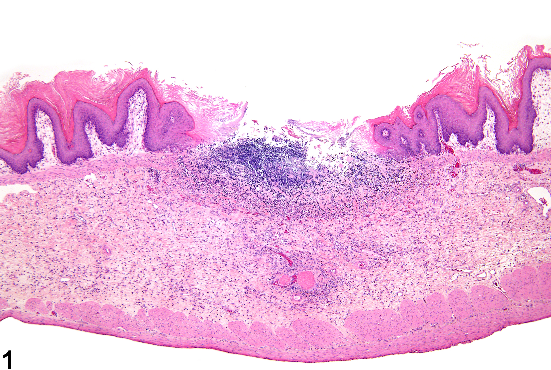
Stomach, Forestomach - Ulcer in a male F344/N rat from a chronic study. A portion of the epithelium is absent, exposing the underlying submucosa.
All Images

Stomach, Forestomach - Ulcer in a male F344/N rat from a chronic study. A portion of the epithelium is absent, exposing the underlying submucosa.

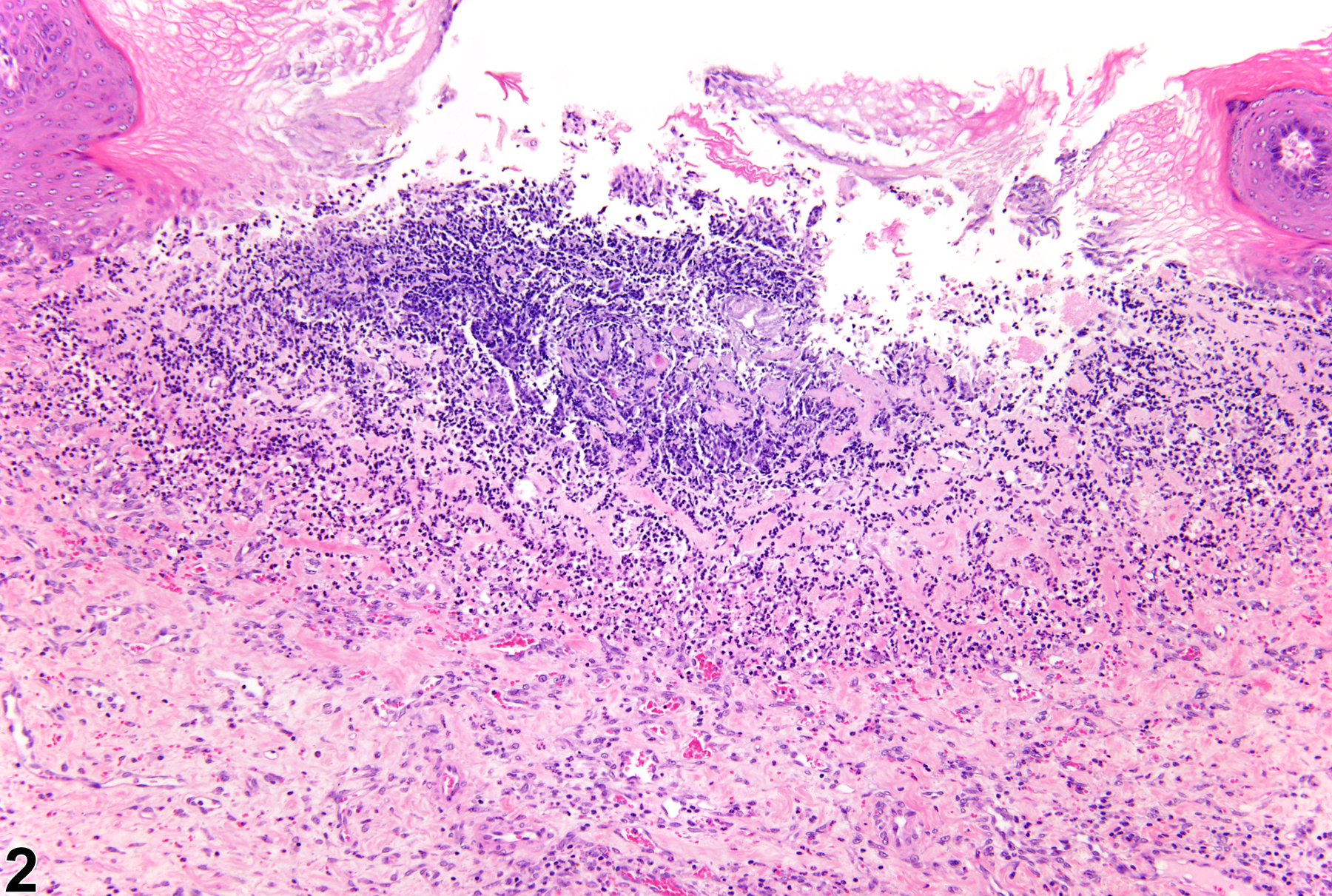
Stomach, Forestomach - Ulcer in a male F344/N rat from a chronic study (higher magnification of Figure 1). There is some inflammation associated with the ulcer.

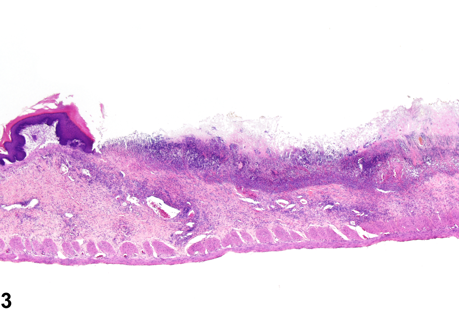
Stomach, Forestomach - Ulcer in a male F344/N rat from a chronic study. A large portion of the epithelium is absent, exposing the underlying submucosa.

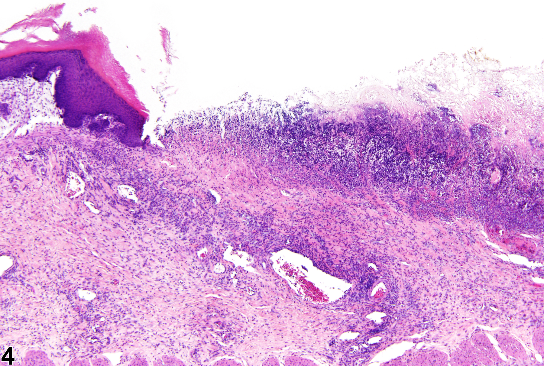
Stomach, Forestomach - Ulcer in a male F344/N rat from a chronic study (higher magnification of Figure 3). A significant amount inflammation is associated with the ulcer, as well as hyperplasia of the adjacent epithelium.

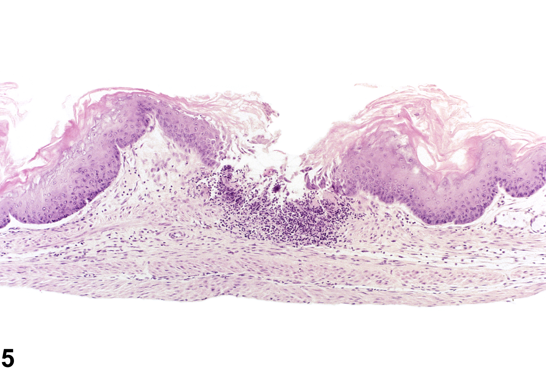
Stomach, Forestomach - Ulcer in a male B6C3F1 mouse from a subchronic study. A large portion of the epithelium is absent, exposing the underlying submucosa.