U.S. flag

An official website of the United States government

Dot gov

The .gov means it's official.
Federal government websites often end in .gov or .mil. Before sharing sensitive information, make sure you're on a federal government site.

Https

The site is secure.
The https:// ensures that you are connecting to the official website and that any information you provide is encrypted and transmitted securely.

Skip to Main Navigation
Skip to Page Content
Skip to Atlas Navigation

Stomach, Glandular Stomach - Amyloid

Image of amyloid in the glandular stomach from a female Swiss Webster mouse in a chronic study
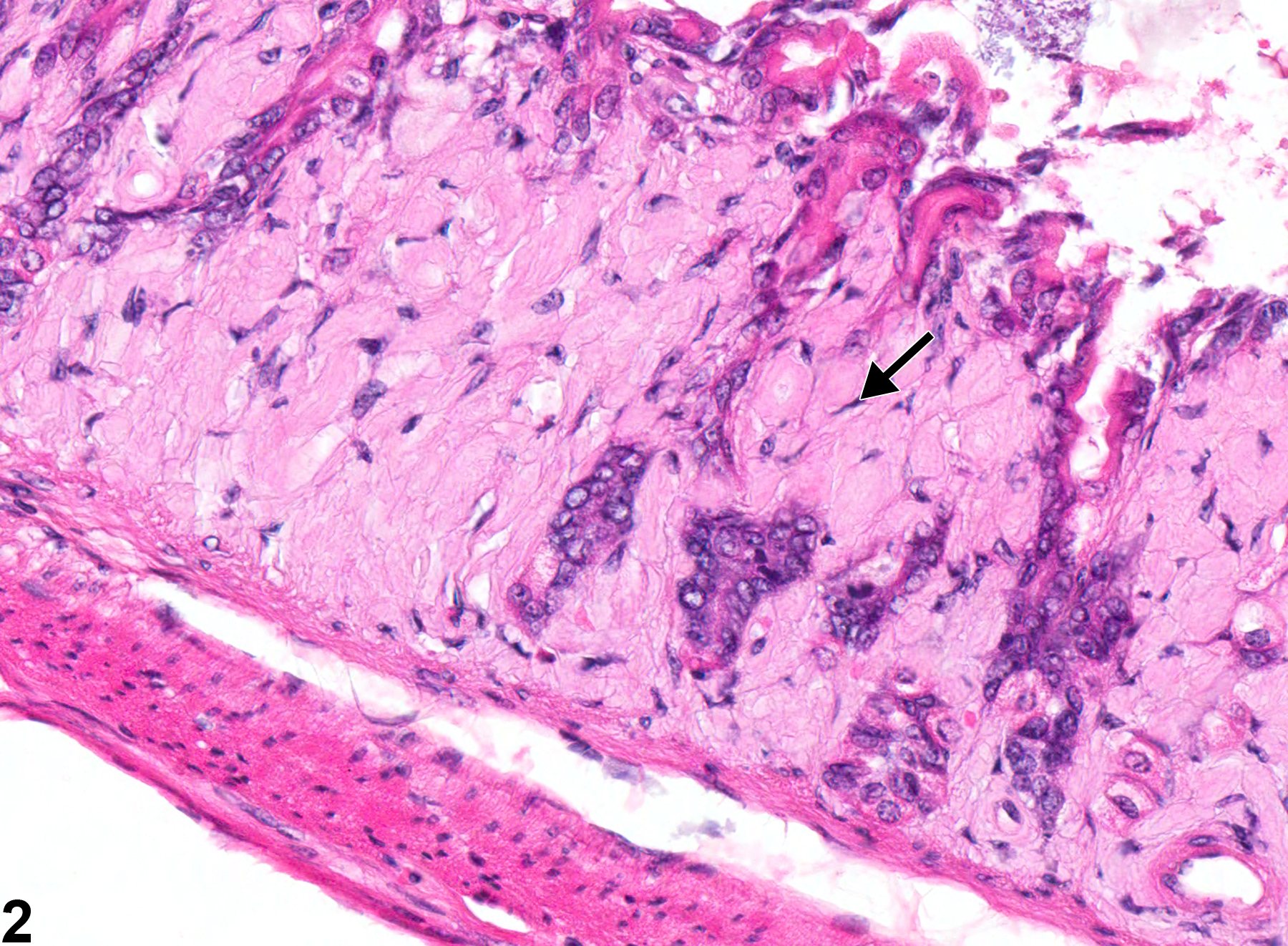
Stomach, Glandular stomach - Amyloid in a female Swiss Webster mouse from a chronic study. Amyloid (arrow) is deposited in the lamina propria.
Figure 1 of 2
Image of amyloid in the glandular stomach from a female Swiss Webster mouse in a chronic study
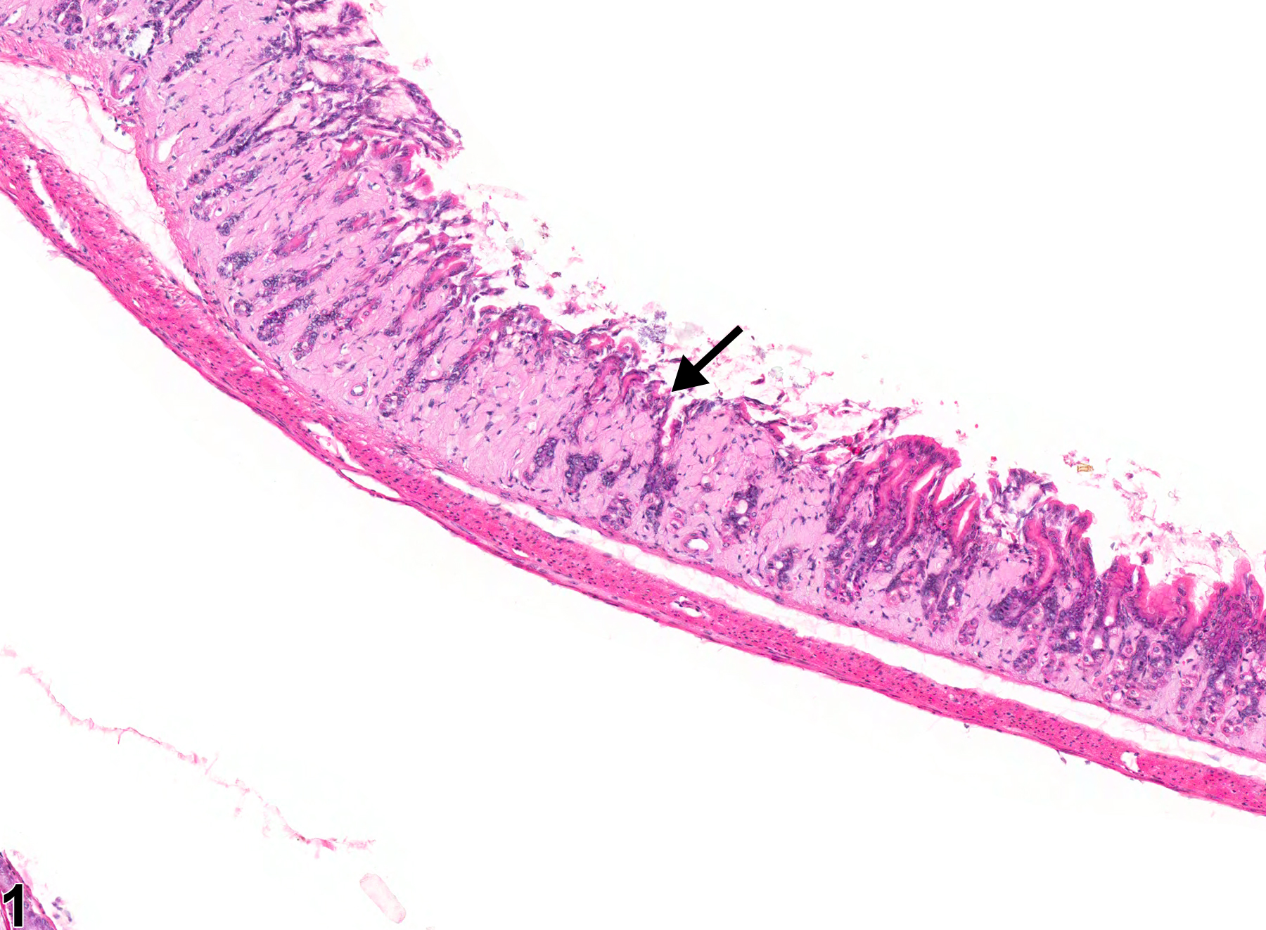
Stomach, Glandular stomach - Amyloid (arrow) in a female Swiss Webster mouse from a chronic study (higher magnification of Figure 1). The deposition of amyloid in the lamina propria results in the loss of glandular epithelial cells.
Figure 2 of 2
next arrow

comment:

Amyloid deposition (amyloidosis) is a systemic disease that is rare in B6C3F1 BALB/c, and C3H mice but common in CD-1, A, Swiss Webster, SJL, and B6 mice and can be a cause of death. The incidence can be increased in association with fighting among group-housed males and with ectoparasitism. Amyloid appears as an amorphous, eosinophilic, hyaline, extracellular substance that, with progressive accumulation, results in pressure atrophy of adjacent cells and tissue. Generalized (systemic) amyloid deposition can be seen in many tissues, including gastrointestinal tract, spleen, liver, kidney, tongue, and mesenteric lymph node. Amyloid deposition in the glandular stomach ( Figure 1image opens in a pop-up window and Figure 2image opens in a pop-up window ) generally occurs in the lamina propria and submucosa. Amyloid deposits in the glomeruli of the kidney are usually the cause of death in animals that die with amyloidosis. Congo red stains amyloid orange to orange red and under polarized light imparts a light green, so-called apple green fluorescence.

recommendation:

Whenever present, amyloid deposits should be diagnosed as "amyloid" and graded based on the extent of the amyloid deposits. The loss of cells secondary to amyloid deposition should not be diagnosed separately but should be described in the narrative.

references:

Bucher JR, Shackelford CC, Haseman JK, Johnson JD, Kurtz PJ, Persing RL. 1994. Carcinogenicity studies of oxazepam in mice. Fundam Appl Toxicol 23:280-297.
Abstract: http://www.sciencedirect.com/science/article/pii/S0272059084711067

Engelhardt JA, Gries CL, Long GG. 1993. Incidence of spontaneous neoplastic and nonneoplastic lesions in Charles River CD-1 mice varies with the breeding origin. Toxicol Pathol 21:538-541.
Full Text: http://tpx.sagepub.com/content/21/6/538.full.pdf

Frith CH, Chandra M. 1991. Incidence, distribution, and morphology of amyloidosis in Charles Rivers CD-1 mice. Toxicol Pathol 19:123-127.
Full Text: http://tpx.sagepub.com/content/19/2/123.full.pdf

Frith CH, Goodman DG, Boysen BG. 2007. The mouse. In: Animal Models in Toxicology, 2nd ed (Gad SC, ed). CRC Press, Boca Raton, FL, 19-146.
Abstract: http://www.crcnetbase.com/isbn/9781420014204

Greaves P. 2007. Digestive system. In: Histopathology of Preclinical Toxicity Studies, 3rd ed. Academic Press, London, 334-456.
Abstract: http://www.sciencedirect.com/science/book/9780444527714

Myers RK, McGavin MD. 2007. Cellular and tissue responses to injury. In: Pathologic Basis of Veterinary Disease, 4th ed (McGavin MD, Zachary JF, eds). Mosby, St Louis, MO, 3-62.

Percy DH, Barthold SW. 2001. Mouse. In: Pathology of Laboratory Rodents and Rabbits, 2nd ed. Iowa State Press, Ames, 3-106.

Ward JM, Mann PC, Morishima H, Frith CH. 1999. Thymus, spleen, and lymph nodes. In: Pathology of the Mouse (Maronpot RR, ed). Cache River Press, St Louis, MO, 333-456.
Abstract: http://www.cacheriverpress.com/books/pathmouse.htm