

Alimentary System
Stomach, Glandular Stomach, Glands - Cyst
Narrative
Bertram TA, Markovits JE, Juliana MM. 1996. Non-proliferative lesions of the alimentary canal in rats GI-1. In Guides for Toxicologic Pathology. STP/ARP/AFIP, Washington, DC, 1-16.
Full Text: https://www.toxpath.org/docs/SSNDC/GINonproliferativeRat.pdfBrown HR, Hardisty JF. 1990. Oral cavity, esophagus and stomach. In: Pathology of the Fischer Rat (Boorman GA, Montgomery CA, MacKenzie WF, eds). Academic Press, San Diego, CA, 9-30.
Abstract: https://www.ncbi.nlm.nih.gov/nlmcatalog/9002563Frantz JD, Betton GR, Cartwright ME, Crissman JW, Macklin AW, Maronpot RR. 1991. Proliferative lesions of the non-glandular and glandular stomach in rats GI-3. In Guides for Toxicologic Pathology. STP/ARP/AFIP, Washington, DC, 1-20.
Full Text: https://www.toxpath.org/docs/SSNDC/StomachProliferativeRat.pdfNational Toxicology Program. 1988. NTP TR-336. Toxicology and Carcinogenesis Studies of Penicillin VK (CAS No. 132-98-9) in F344/N Rats and B6C3F1 Mice (Gavage Studies). NTP, Research Triangle Park, NC.
Abstract: https://ntp.niehs.nih.gov/go/8907National Toxicology Program. 2000. NTP TR-491. Toxicology and Carcinogenesis Studies of Methyleugenol (CAS No. 93-15-2) in F344/N Rats and B6C3F1 Mice (Gavage Studies). NTP, Research Triangle Park, NC.
Abstract: https://ntp.niehs.nih.gov/go/10172National Toxicology Program. 2010. NTP TR-558. Toxicology and Carcinogenesis Studies of 3,3’,4,4’-Tetrachloroazobenzene (TCAB) (CAS No. 14047-09-7) in Harlan Sprague Dawley Rats and B6C3F1 Mice (Gavage Studies). NTP, Research Triangle Park, NC.
Abstract: https://ntp.niehs.nih.gov/go/33564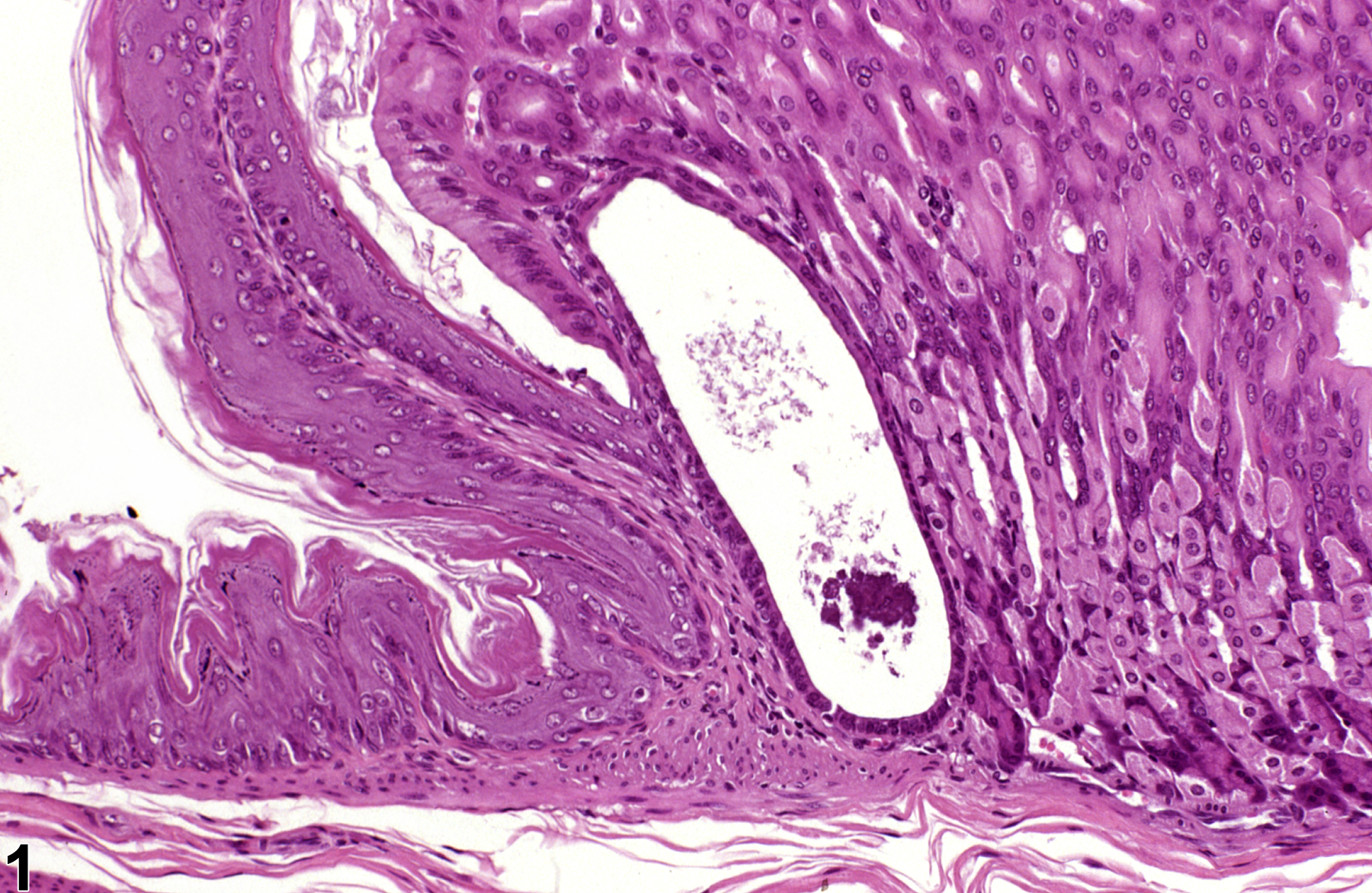
Stomach, Glandular stomach, Glands - Cyst in a male B6C3F1 mouse from a subchronic study. This cystic gland is lined by slightly flattened epithelium.
All Images

Stomach, Glandular stomach, Glands - Cyst in a male B6C3F1 mouse from a subchronic study. This cystic gland is lined by slightly flattened epithelium.