U.S. flag

An official website of the United States government

Dot gov

The .gov means it's official.
Federal government websites often end in .gov or .mil. Before sharing sensitive information, make sure you're on a federal government site.

Https

The site is secure.
The https:// ensures that you are connecting to the official website and that any information you provide is encrypted and transmitted securely.

Skip to Main Navigation
Skip to Page Content
Skip to Atlas Navigation

Stomach, Glandular Stomach - Ectopic Tissue

Image of ectopic tissue (pancreas) in the glandular stomach from a female B6C3F1 mouse in a chronic study
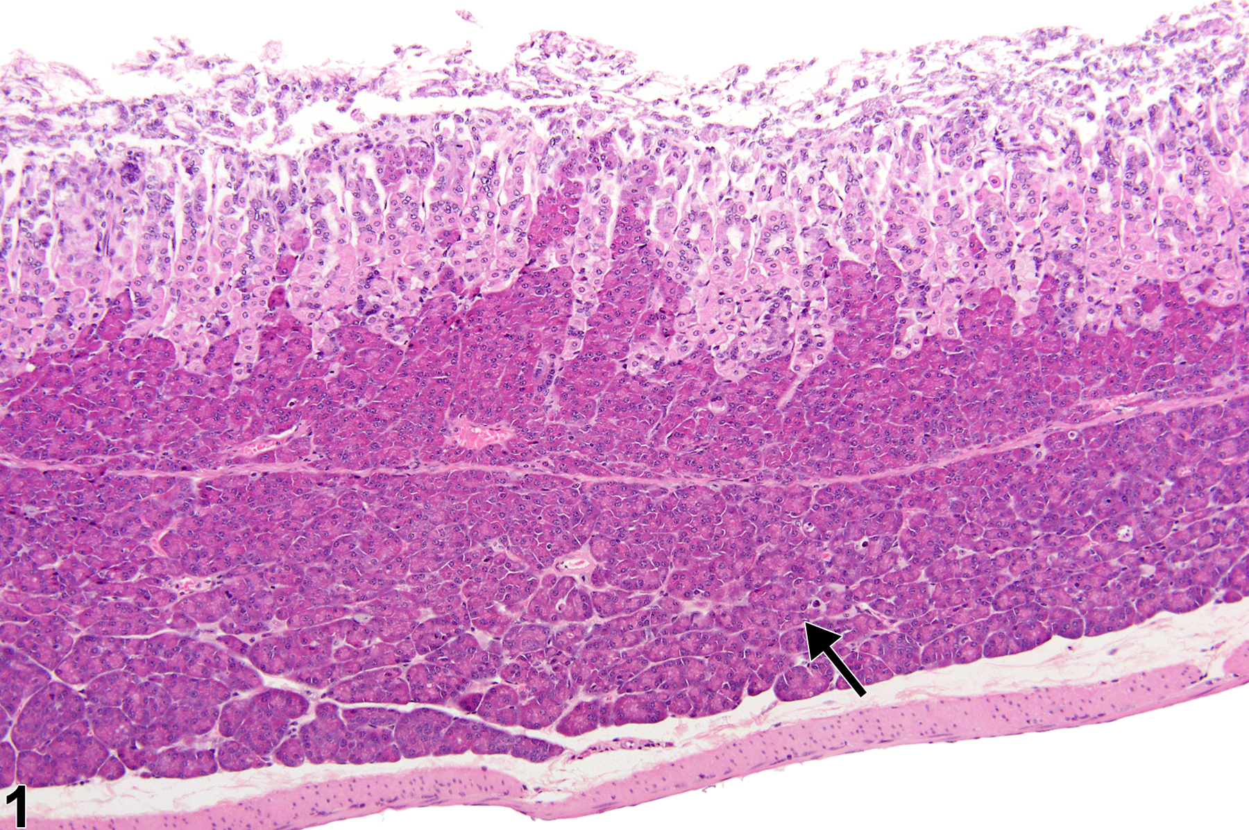
Stomach, Glandular stomach - Ectopic tissue, Pancreas in a female B6C3F1 mouse from a chronic study. There are pancreatic acinar cells in submucosa of the glandular stomach (arrow).
Figure 1 of 6
Image of ectopic tissue (pancreas) in the glandular stomach from a female B6C3F1 mouse in a chronic study
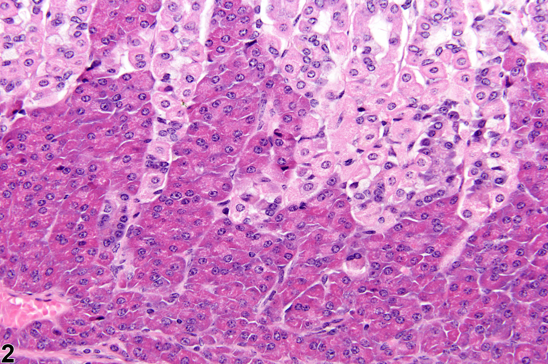
Stomach, Glandular stomach - Ectopic tissue, Pancreas in a female B6C3F1 mouse from a chronic study (higher magnification of Figure 1). There are pancreatic acinar cells in the lamina propria of the glandular stomach that extend into the lamina propria.
Figure 2 of 6
Image of ectopic tissue (liver) in the glandular stomach from a female B6C3F1 mouse in a chronic study
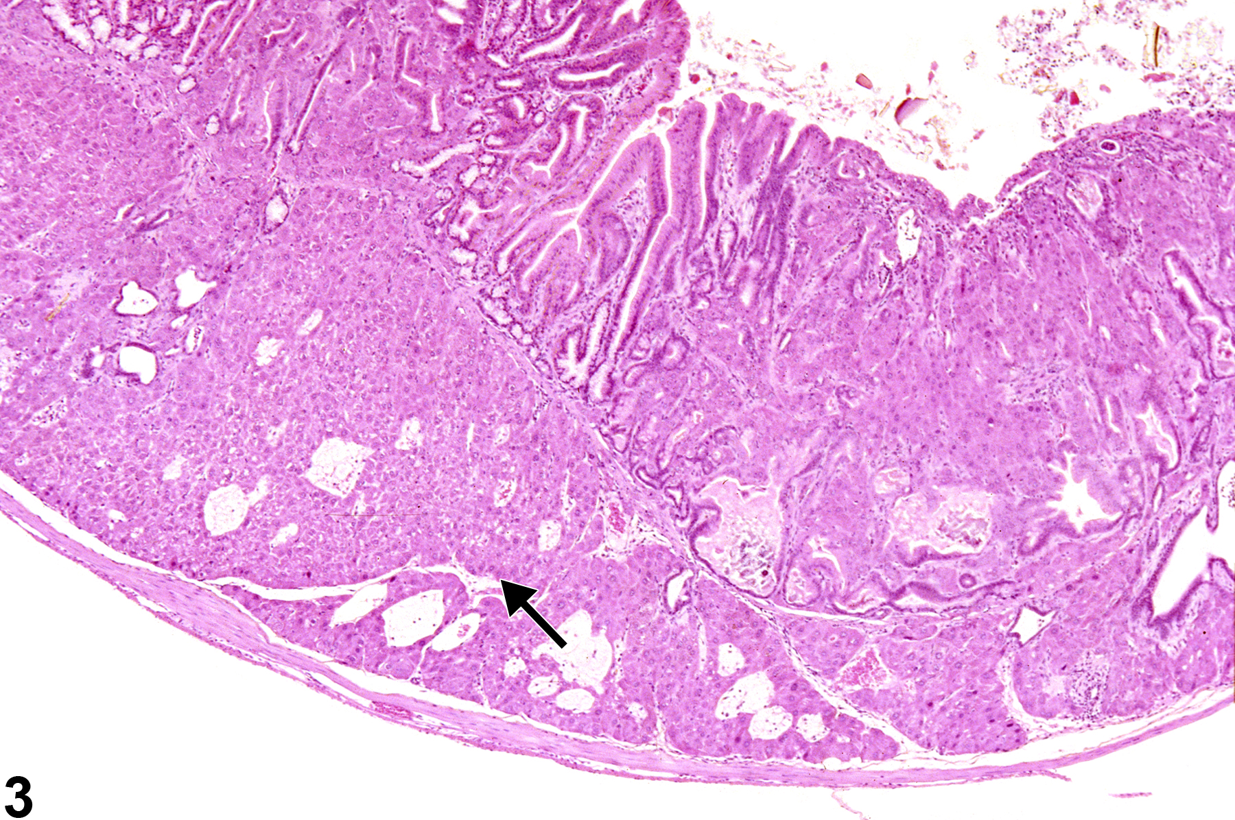
Stomach, Glandular stomach - Ectopic tissue, Liver in a female B6C3F1 mouse from a chronic study. There are hepatocytes in submucosa of the glandular stomach (arrow).
Figure 3 of 6
Image of ectopic tissue (liver) in the glandular stomach from a female B6C3F1 mouse in a chronic study
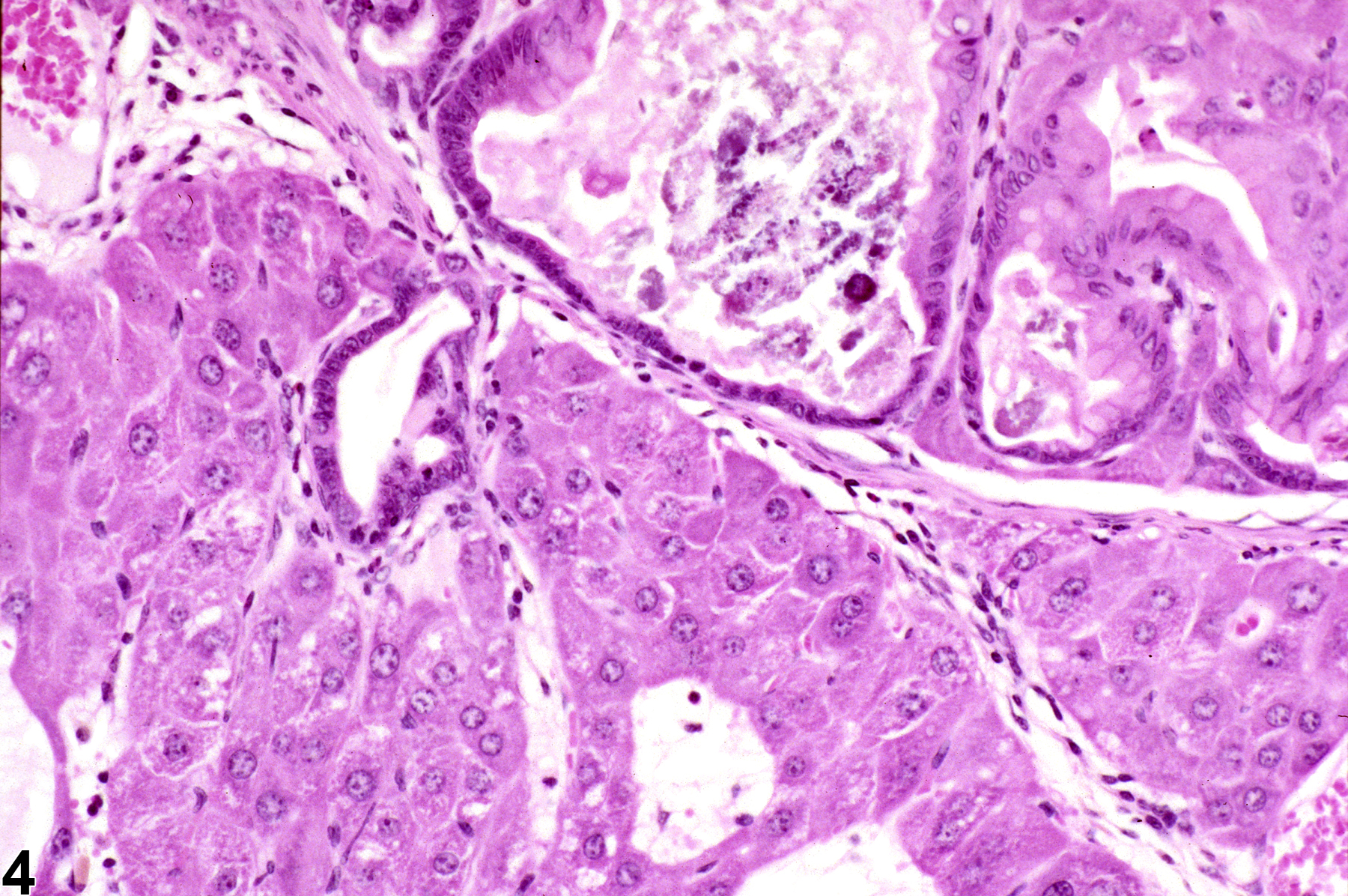
Stomach, Glandular stomach - Ectopic tissue, Liver in a female B6C3F1 mouse from a chronic study (higher magnification of Figure 3). There are hepatocytes in submucosa of the glandular stomach.
Figure 4 of 6
Image of ectopic tissue (intestine) in the glandular stomach from a male F344/N rat in a chronic study
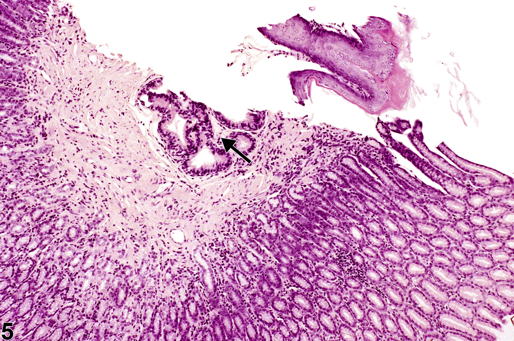
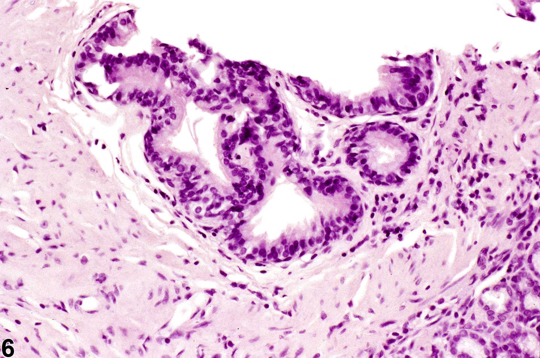
Stomach, Glandular stomach - Ectopic tissue, Intestine in a male F344/N rat from a chronic study. There is intestinal epithelial tissue in the submucosa of the glandular stomach (arrow).
Figure 5 of 6
Image of ectopic tissue (intestine) in the glandular stomach from a male F344/N rat in a chronic study
Stomach, Glandular stomach - Ectopic tissue, Intestine in a male F344/N rat from a chronic study (higher magnification of Figure 3). There is intestinal epithelial tissue in the submucosa of the glandular stomach.
Figure 6 of 6
next arrow

comment:

Ectopic pancreas ( Figure 1image opens in a pop-up window and Figure 2image opens in a pop-up window ), liver ( Figure 3image opens in a pop-up window and Figure 4image opens in a pop-up window ) or intestines ( Figure 5image opens in a pop-up window and Figure 6image opens in a pop-up window ) occur rarely in the submucosa of the glandular stomach. Hepatocytes in the submucosa/lamina propria of the glandular stomach have been reported as a rare occurrence in the B6C3F1 mouse, as well as other mouse strains. Mice from control and treatment groups and both sexes have been found with gastric hepatocytes. This condition may arise from metaplasia of a primary stomach cell type but more likely is the result of a developmental defect and is considered an incidental finding. Invasion or metastasis from a liver neoplasm must be ruled out in all cases, even if the cells do not appear neoplastic. Ectopic pancreatic tissue, composed of acinar and ductal cells, has also been described in the submucosa of the mouse stomach. Foci of intestinal tissue can occur in the glandular stomach ( Figure 5image opens in a pop-up window and Figure 6image opens in a pop-up window ). This is considered an incidental congenital lesion similar to hepatic/pancreatic ectopic foci in the stomach. It is differentiated from intestinal metaplasia by occurrence outside of the mucosal epithelium and a lack of association with other changes in the epithelium, such as hyperplasia, atypical changes (dysplasia), or gastric carcinoma.

recommendation:

Whenever present, ectopic tissue should be diagnosed but not graded. The diagnosis should be modified with the type of ectopic tissue.

references:

Brown HR, Hardisty JF. 1990. Oral cavity, esophagus and stomach. In: Pathology of the Fischer Rat (Boorman GA, Montgomery CA, MacKenzie WF, eds). Academic Press, San Diego, CA, 9-30.
Abstract: https://www.ncbi.nlm.nih.gov/nlmcatalog/9002563

Greaves P. 2007. Digestive system. In: Histopathology of Preclinical Toxicity Studies, 3rd ed. Academic Press, London, 334-456.
Abstract: http://www.sciencedirect.com/science/book/9780444527714

Leininger JR, Jokinen MP, Dangler CA, Whiteley LO. 1999. Oral cavity, esophagus, and stomach. In: Pathology of the Mouse (Maronpot RR, ed). Cache River Press, St Louis, MO, 29-48.
Abstract: http://www.cacheriverpress.com/books/pathmouse.htm

Leininger JR, McDonald MM, Abbott DP. 1990. Hepatocytes in the mouse stomach. Toxicol Pathol 18:678-686.
Abstract: https://www.ncbi.nlm.nih.gov/pubmed/2093226

Maekawa A, Enomoto M, Hirouchi Y, Yamakawa S. 1996. Changes in the upper digestive tract and stomach. In: Pathobiology of the Aging Mouse, Vol 2 (Mohr U, Dungworth DL, Capen CC, Carlton WW, Sundberg JP, Ward JM, eds). ILSI Press, Washington, DC, 267-286.

Yamakawa S, Iwata H, Hirouchi Y, Inomoto M. 1993. Hepatic metaplasia in glandular stomach and pancreas of mice. Toxicol Pathol 21:590.