

Alimentary System
Stomach, Glandular Stomach - Edema
Narrative
Hirose M, Hakoi K, Takahashi S, Hoshiya T, Akagi K, Lin C, Saito K, Kaneko H, Shirai T. 1999. Sequential morphological and biological changes in the glandular stomach induced by oral administration of catechol to male F344 rats. Toxicol Pathol 27:448-455.
Abstract: https://www.ncbi.nlm.nih.gov/pubmed/10485826Mosier, DA. 2007. Vascular disorders and thrombosis. In: Pathologic Basis of Veterinary Disease, 4th ed (McGavin MD, Zachary JF, eds). Mosby, St Louis, MO, 63-99.
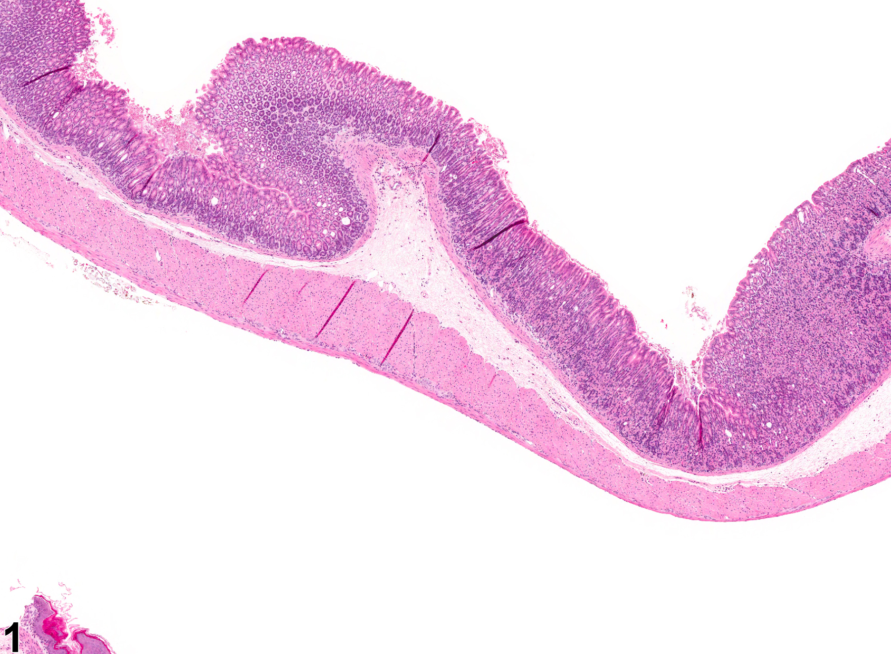
Stomach, Glandular stomach - Edema in a female B6C3F1 mouse from a chronic study. The submucosa is expanded by fluid.
All Images

Stomach, Glandular stomach - Edema in a female B6C3F1 mouse from a chronic study. The submucosa is expanded by fluid.
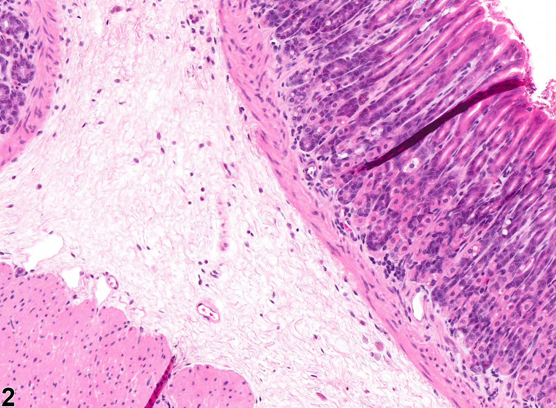
Stomach, Glandular stomach - Edema in a female B6C3F1 mouse from a chronic study (higher magnification of Figure 1). The submucosa is expanded by fluid.
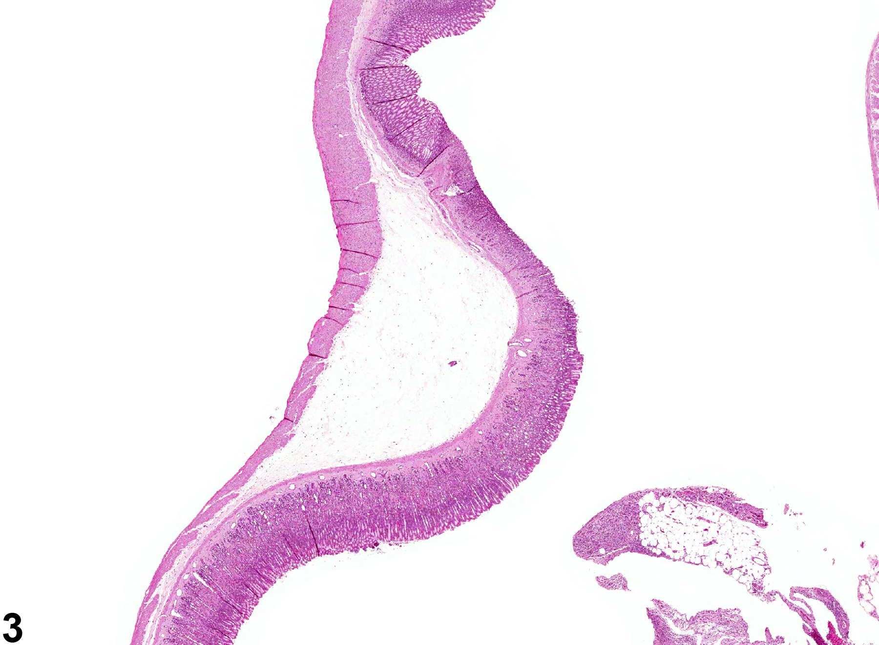
Stomach, Glandular stomach - Edema in a female B6C3F1 mouse from a chronic study (higher magnification of Figure 1). The submucosa is expanded by fluid.
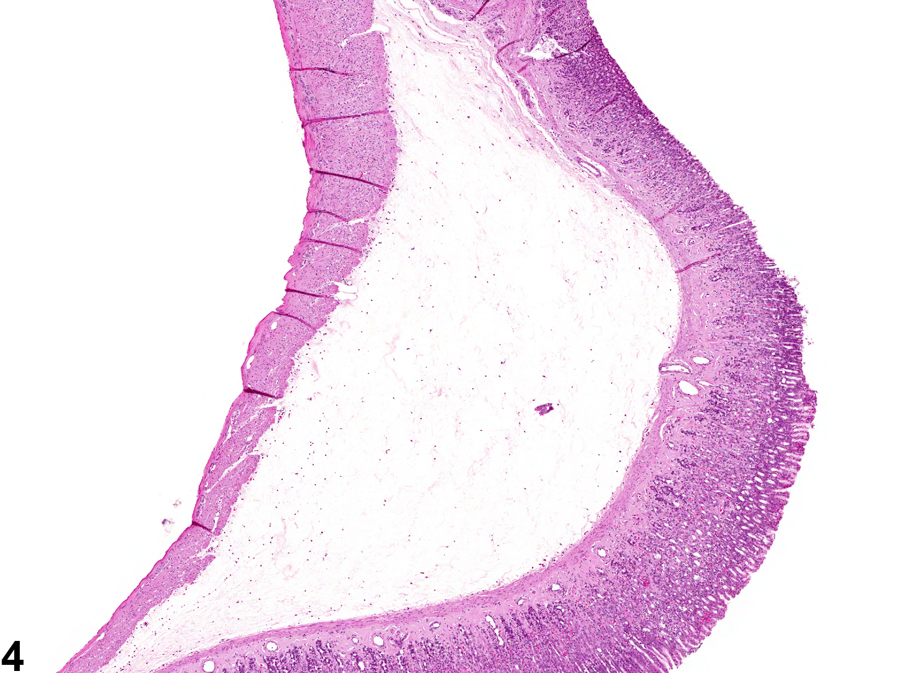
Stomach, Glandular stomach - Edema in a female B6C3F1 mouse from a chronic study (higher magnification of Figure 1). The submucosa is expanded by fluid.