

Alimentary System
Stomach, Glandular Stomach - Erosion
Narrative
Ackerman SH, Hofer MA, Weiner H. 1975. Age at maternal separation and gastric erosion susceptibility in the rat. Psychosom Med 37:180-183.
Abstract: https://www.ncbi.nlm.nih.gov/pubmed/1079604Betton GR. 1998. The digestive system I: The gastrointestinal tract and exocrine pancreas. In: Target Organ Pathology (Turton J, Hooson J, eds). Taylor and Francis, London, 29-60.
Brown HR, Hardisty JF. 1990. Oral cavity, esophagus and stomach. In: Pathology of the Fischer Rat (Boorman GA, Montgomery CA, MacKenzie WF, eds). Academic Press, San Diego, CA, 9-30.
Abstract: https://www.ncbi.nlm.nih.gov/nlmcatalog/9002563Hirose M, Hakoi K, Takahashi S, Hoshiya T, Akagi K, Lin C, Saito K, Kaneko H, Shirai T. 1999. Sequential morphological and biological changes in the glandular stomach induced by oral administration of catechol to male F344 rats. Toxicol Pathol 27:448-455.
Abstract: https://www.ncbi.nlm.nih.gov/pubmed/10485826Leininger JR, Jokinen MP, Dangler CA, Whiteley LO. 1999. Oral cavity, esophagus, and stomach. In: Pathology of the Mouse (Maronpot RR, ed). Cache River Press, St Louis, MO, 29-48.
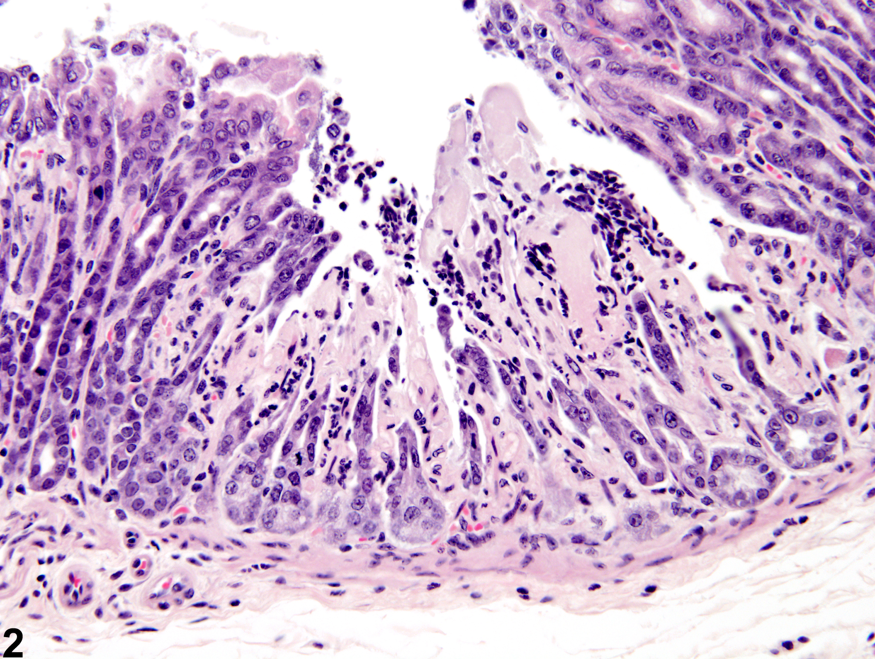
Stomach, Glandular stomach - Erosion in male B6C3F1 mouse from a subchronic study. The lesion does not extend through the entire mucosa.
All Images

Stomach, Glandular stomach - Erosion in male B6C3F1 mouse from a subchronic study. The lesion does not extend through the entire mucosa.

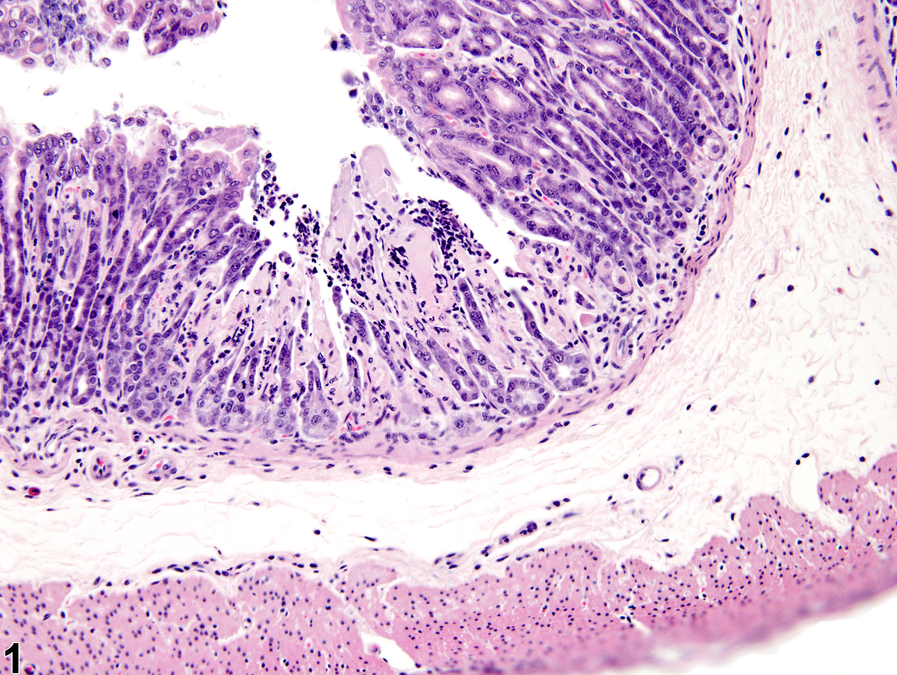
Stomach, Glandular stomach - Erosion in male B6C3F1 mouse from a subchronic study (higher magnification of Figure 1). The lesion does not extend through the entire mucosa.