U.S. flag

An official website of the United States government

Dot gov

The .gov means it's official.
Federal government websites often end in .gov or .mil. Before sharing sensitive information, make sure you're on a federal government site.

Https

The site is secure.
The https:// ensures that you are connecting to the official website and that any information you provide is encrypted and transmitted securely.

Skip to Main Navigation
Skip to Page Content
Skip to Atlas Navigation

Stomach, Glandular Stomach - Foreign body

Image of foreign body in the glandular stomach from a male B6C3F1 mouse in a chronic study
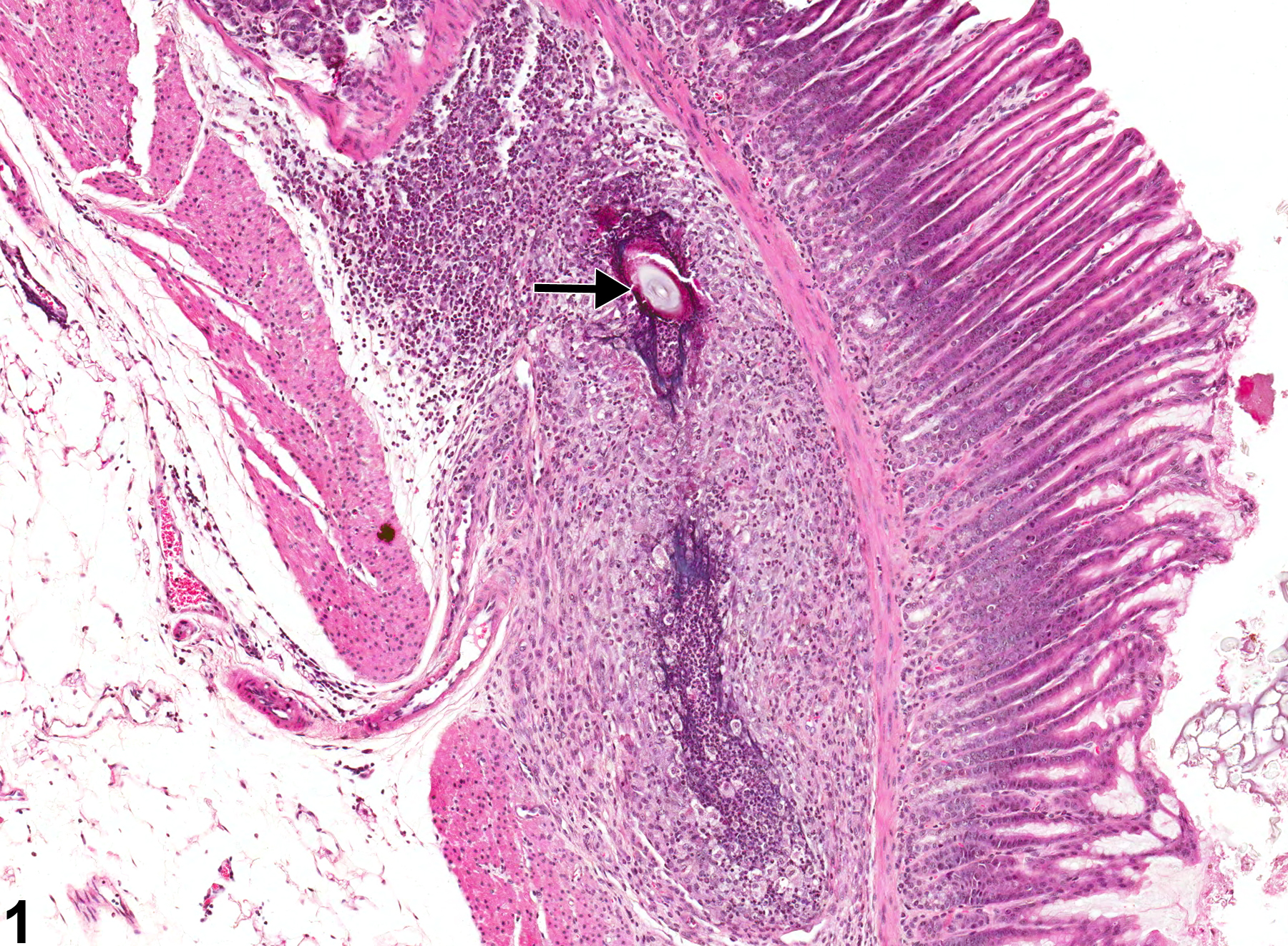
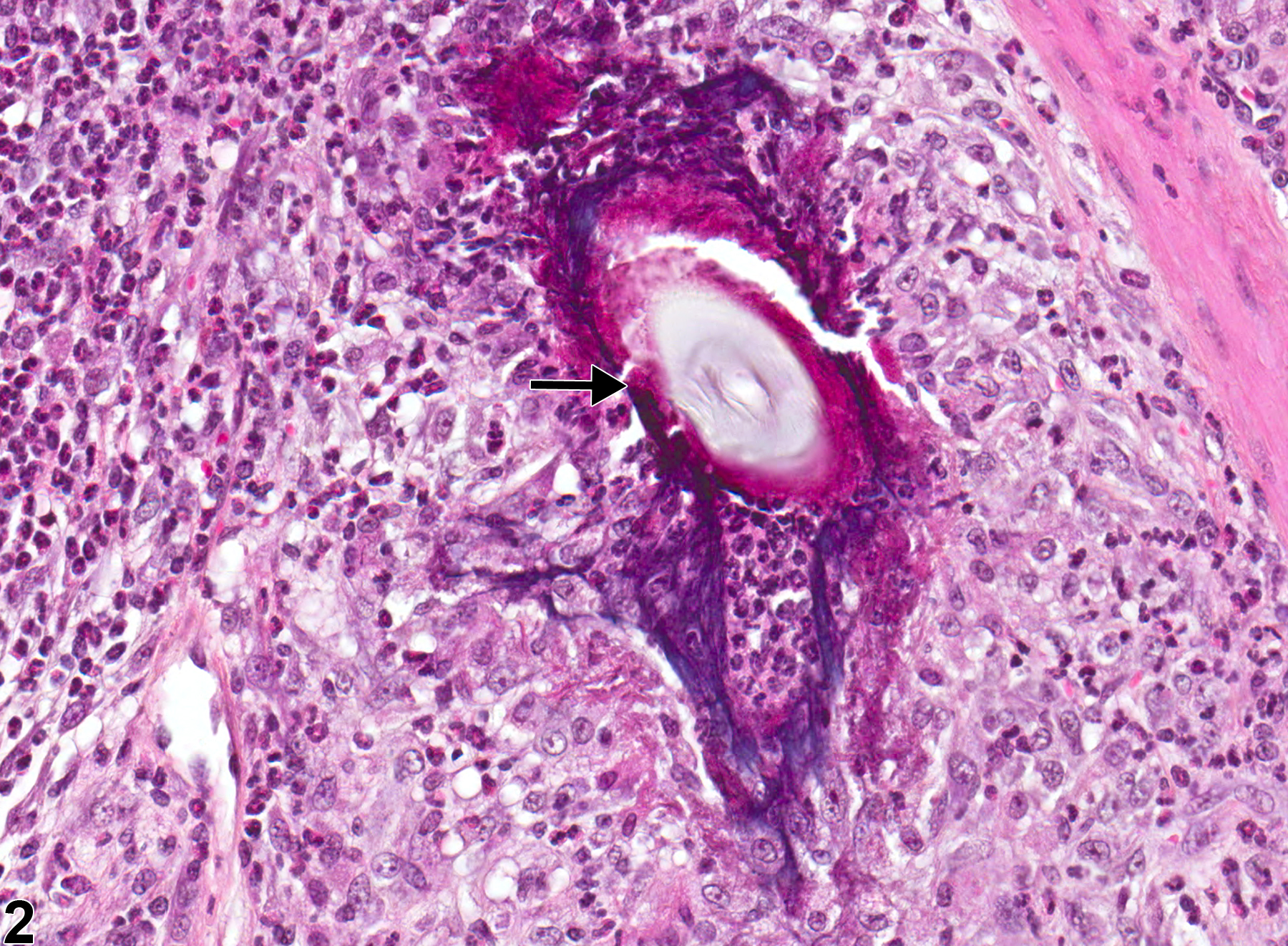
Stomach, Glandular stomach - Foreign body in a male B6C3F1 mouse from a chronic study. There is a foreign body (arrow) in the submucosa with secondary inflammation.
Figure 1 of 2
Image of foreign body in the glandular stomach from a male B6C3F1 mouse in a chronic study
Stomach, Glandular stomach - Foreign body in a male B6C3F1 mouse from a chronic study (higher magnification of Figure 1). There is a foreign body (arrow) in the submucosa with secondary inflammation.
Figure 2 of 2
next arrow

comment:

Hair ( Figure 1image opens in a pop-up window and Figure 2image opens in a pop-up window , arrows) or feed material can become embedded in the wall of the glandular stomach secondary to injury from a gavage procedure or ulceration from xenobiotics. The process of ulceration exposes the underlying propria and muscular wall to gastric contents and allows hair and food material to penetrate into the granulation tissue, which forms beneath the ulcer.

recommendation:

A primary foreign body should be diagnosed but not graded. If a foreign body causing a significant inflammatory response, then both the foreign body and inflammation are diagnosed. In the case illustrated above, both the foreign body (hair) and the inflammation (granulomatous) should be diagnosed. A foreign body such as hair or feed material that is passively adhered to an ulcerated surface should not be diagnosed separately; it can be described in the narrative.

references:

Brown HR, Hardisty JF. 1990. Oral cavity, esophagus and stomach. In: Pathology of the Fischer Rat (Boorman GA, Montgomery CA, MacKenzie WF, eds). Academic Press, San Diego, CA, 9-30.
Abstract: https://www.ncbi.nlm.nih.gov/nlmcatalog/9002563

Deardorff TL, Kliks MM, Desowitz RS. 1983. Histopathology induced by larval Terranova (type HA) (Nematoda: Anisakinae) in experimentally infected rats. J Parasitol 69:191-195.
Abstract: https://www.ncbi.nlm.nih.gov/pubmed/6827436

Leininger JR, Jokinen MP, Dangler CA, Whiteley LO. 1999. Oral cavity, esophagus, and stomach. In: Pathology of the Mouse (Maronpot RR, ed). Cache River Press, St Louis, MO, 29-48.
Abstract: http://www.cacheriverpress.com/books/pathmouse.htm