

Alimentary System
Stomach, Glandular Stomach - Hemorrhage
Narrative
MacKenzie WF, Alison R. 1990. Heart. In: Pathology of the Fischer Rat (Boorman GA, Montgomery CA, MacKenzie WF, eds). Academic Press, San Diego, CA, 461-472.
Abstract: https://www.ncbi.nlm.nih.gov/nlmcatalog/9002563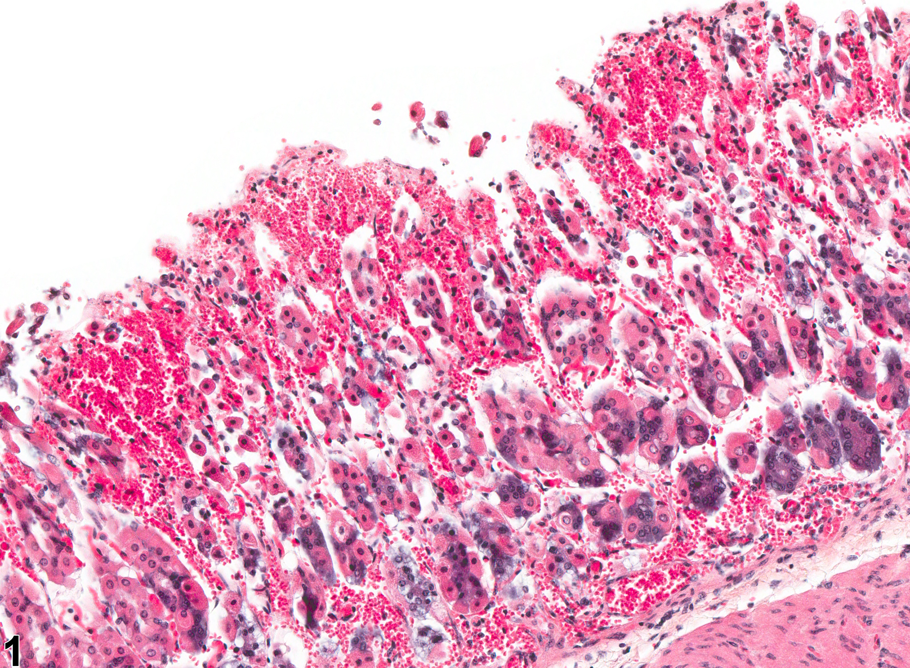
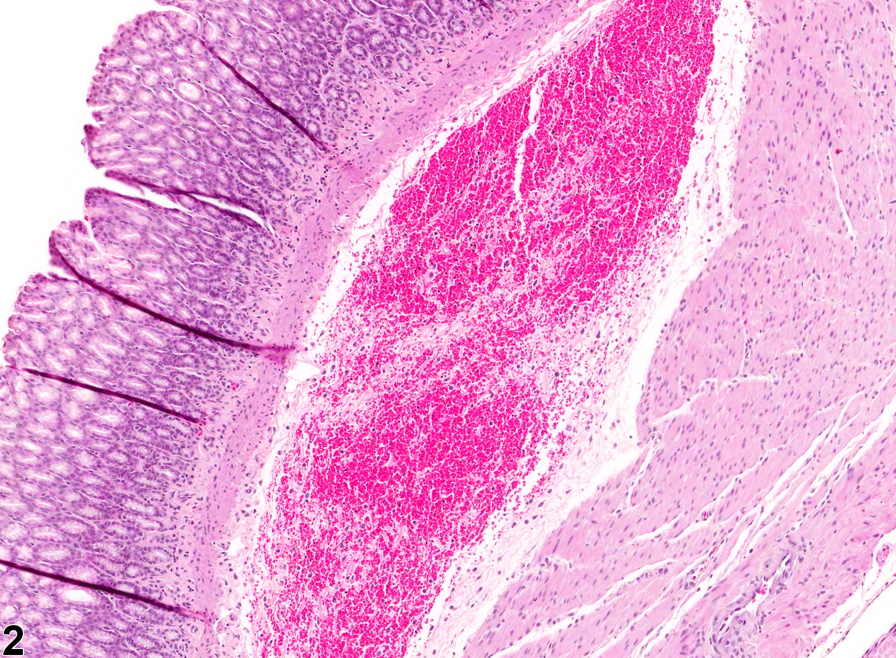
Stomach, Glandular stomach - Hemorrhage in a female B6C3F1 mouse from a subchronic study. There is hemorrhage in the mucosa.
All Images

Stomach, Glandular stomach - Hemorrhage in a female B6C3F1 mouse from a subchronic study. There is hemorrhage in the mucosa.

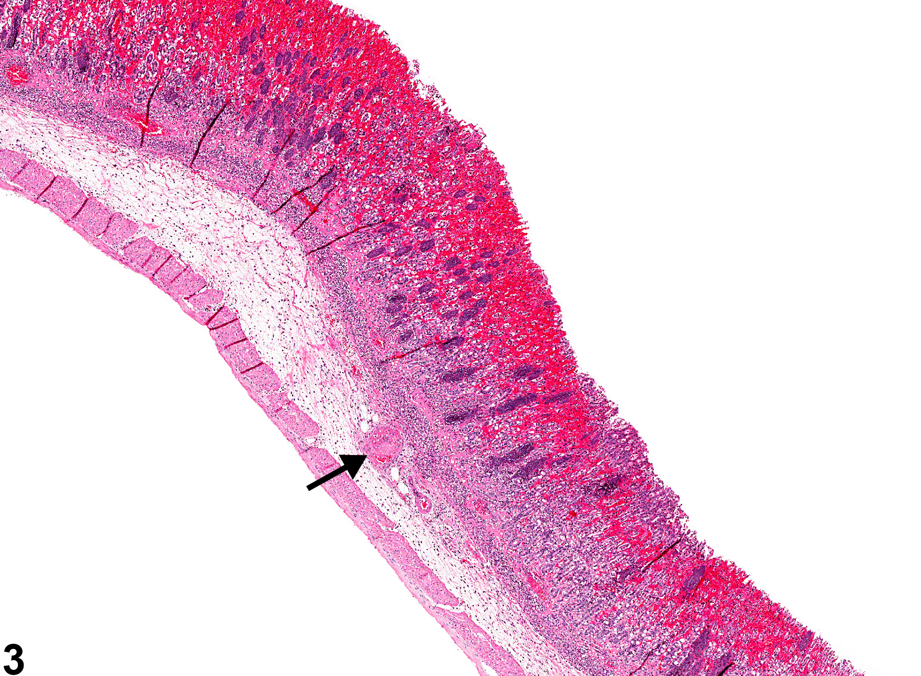
Stomach, Glandular stomach - Hemorrhage in a male F344/N rat from a chronic study. There is hemorrhage in the submucosa.

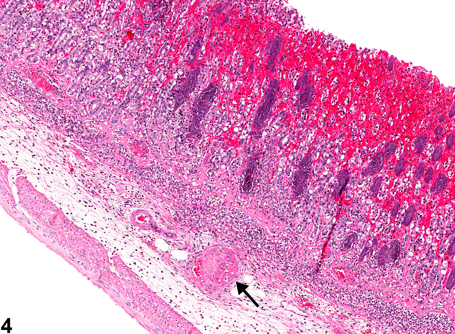
Stomach, Glandular stomach - Hemorrhage in a male F344/N rat from a chronic study. There is hemorrhage in the mucosa and a vascular thrombus in the submucosa (arrow).

Stomach, Glandular stomach - Hemorrhage in a male F344/N rat from a chronic study (higher magnification of Figure 3). There is hemorrhage in the mucosa and a vascular thrombus in the submucosa (arrow).