U.S. flag

An official website of the United States government

Dot gov

The .gov means it's official.
Federal government websites often end in .gov or .mil. Before sharing sensitive information, make sure you're on a federal government site.

Https

The site is secure.
The https:// ensures that you are connecting to the official website and that any information you provide is encrypted and transmitted securely.

Skip to Main Navigation
Skip to Page Content
Skip to Atlas Navigation

Stomach, Glandular Stomach - Mineralization

Image of mineralization in the glandular stomach from a male F344/N rat in a chronic study
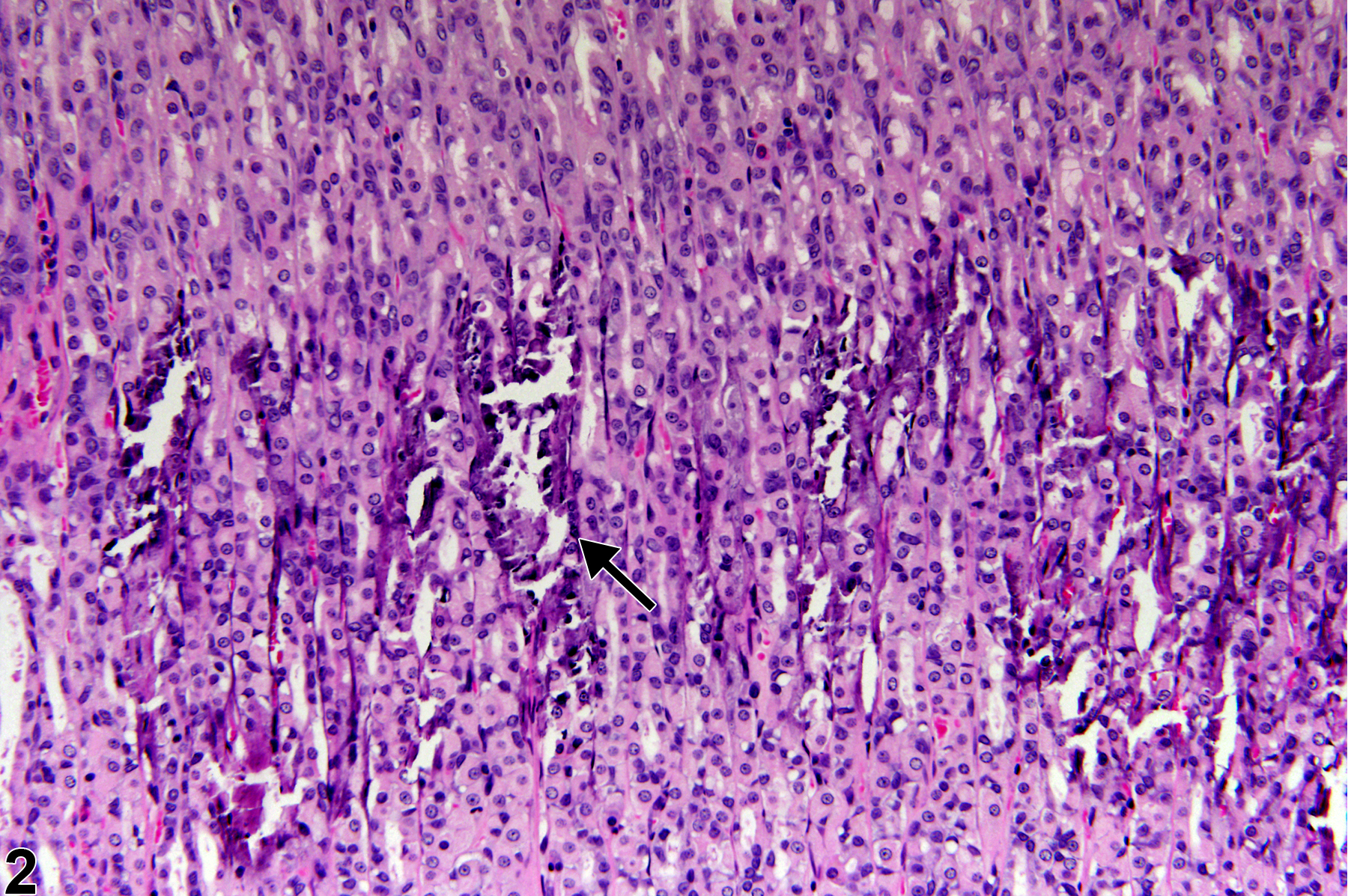
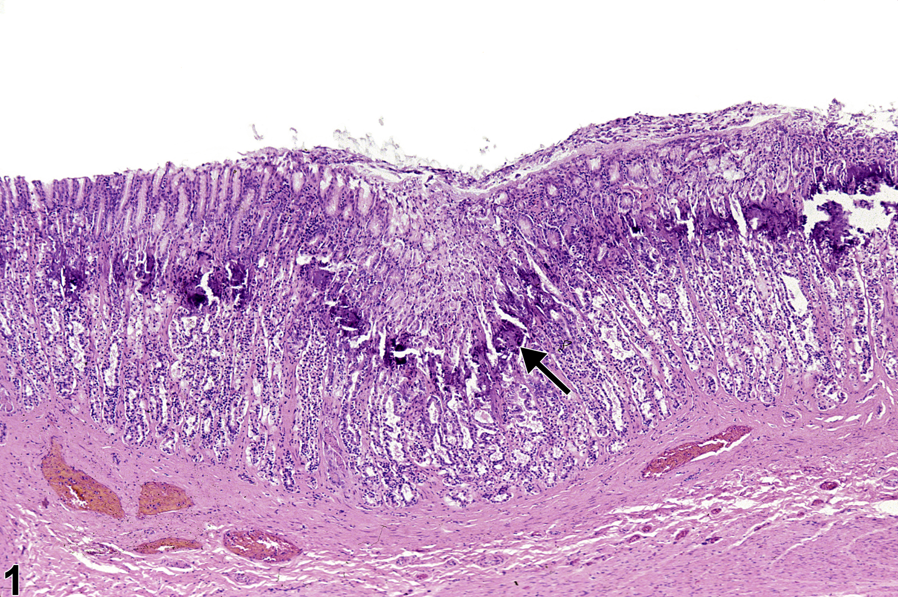
Stomach, Glandular stomach - Mineralization in a male F344/N rat from a chronic study. There is a linear band of mineralization paralleling the parietal rich region in gastric fundus (arrow).
Figure 1 of 2
Image of mineralization in the glandular stomach from a male F344/N rat in a chronic study
Stomach, Glandular stomach - Mineralization in a male F344/N rat from a chronic study (higher magnification of Figure 1). There is a linear band of mineralization paralleling the parietal rich region in gastric fundus (arrow).
Figure 2 of 2
next arrow

comment:

Mineralization characterized by focal aggregates or concretions of densely blue-staining mineral is fairly common in the H&E-stained sections from the stomachs of aged rats, where they are associated with cystic dilation of the gastric glands; mice and hamsters occasionally show similar changes. Gastric mineralization may become marked in rodents when there is disturbance in mineral metabolism, particularly with chronic progressive renal disease and uremia due to altered calcium-phosphorus metabolism and hyperparathyroidism. These changes are characterized by mineralization of the vessels throughout the gastric fundus, muscular tunics, and interstitium in a band paralleling the parietal-cell-rich region of the mucosa ( Figure 1image opens in a pop-up window and Figure 2image opens in a pop-up window , arrows) but not the cardia, antrum, or pylorus. Mineral deposits develop around basement membranes surrounding epithelium and blood vessels. The lamina propria becomes expanded by edema, and fibroplasia of the interstitium develops. The gastric glands become dilated with swelling and degeneration of parietal cells and atrophy of chief cells. Erosion of the glandular epithelium with hemorrhage occurs. Gastric mineralization is frequently accompanied by mucous cell hyperplasia.

recommendation:

Mineralization should be diagnosed and graded depending on the extent of mineralization. Associated lesions, such as inflammation or degeneration of gastric epithelial cells, that are secondary to the mineralization should not be diagnosed separately but should be described in the narrative.

references:

Bertram TA, Markovits JE, Juliana MM. 1996. Non-proliferative lesions of the alimentary canal in rats GI-1. In Guides for Toxicologic Pathology. STP/ARP/AFIP, Washington, DC, 1-16.
Full Text: https://www.toxpath.org/docs/SSNDC/GINonproliferativeRat.pdf

Brown HR, Hardisty JF. 1990. Oral cavity, esophagus and stomach. In: Pathology of the Fischer Rat (Boorman GA, Montgomery CA, MacKenzie WF, eds). Academic Press, San Diego, CA, 9-30.
Abstract: https://www.ncbi.nlm.nih.gov/nlmcatalog/9002563

Greaves P. 2007. Digestive system. In: Histopathology of Preclinical Toxicity Studies, 3rd ed. Academic Press, London, 334-456.
Abstract: http://www.sciencedirect.com/science/book/9780444527714

Leininger JR, Jokinen MP, Dangler CA, Whiteley LO. 1999. Oral cavity, esophagus, and stomach. In: Pathology of the Mouse (Maronpot RR, ed). Cache River Press, St Louis, MO, 29-48.
Abstract: http://www.cacheriverpress.com/books/pathmouse.htm

Newman SJ, Confer AW, Panciera RJ. 2007. Urinary system. In: Pathologic Basis of Veterinary Disease, 4th ed (McGavin MD, Zachary JF, eds). Mosby, St Louis, MO, 613-691.