

Alimentary System
Stomach, Glandular Stomach, Neuroendocrine Cell - Hyperplasia
Narrative
Betton GR. 1998. The digestive system I: The gastrointestinal tract and exocrine pancreas. In: Target Organ Pathology (Turton J, Hooson J, eds). Taylor and Francis, London, 29-60.
Brown HR, Hardisty JF. 1990. Oral cavity, esophagus and stomach. In: Pathology of the Fischer Rat (Boorman GA, Montgomery CA, MacKenzie WF, eds). Academic Press, San Diego, CA, 9-30.
Abstract: https://www.ncbi.nlm.nih.gov/nlmcatalog/9002563DeLellis RA, Dayal Y. 2007. The neuroendocrine system. In: Histology for Pathologists, 3rd ed (Mills SE, ed). Lippincott Williams and Wilkins, Philadelphia, PA, 1189-1210.
Johnson JD, Ryan MJ, Toft JD II, Graves SW, Hejtmancik MR, Cunningham ML, Herbert R, Abdo KM. 2000. Two-year toxicity and carcinogenicity study of methyleugenol in F344/N rats and B6C3F1 mice. J Agric Food Chem 48:3620-3632.
Abstract: http://pubs.acs.org/doi/abs/10.1021/jf000364aNational Toxicology Program. 2000. NTP TR-491. Toxicology and Carcinogenesis Studies of Methyleugenol (CAS No. 93-15-2) in F344/N Rats and B6C3F1 Mice (Gavage Studies). NTP, Research Triangle Park, NC.
Abstract: https://ntp.niehs.nih.gov/go/10172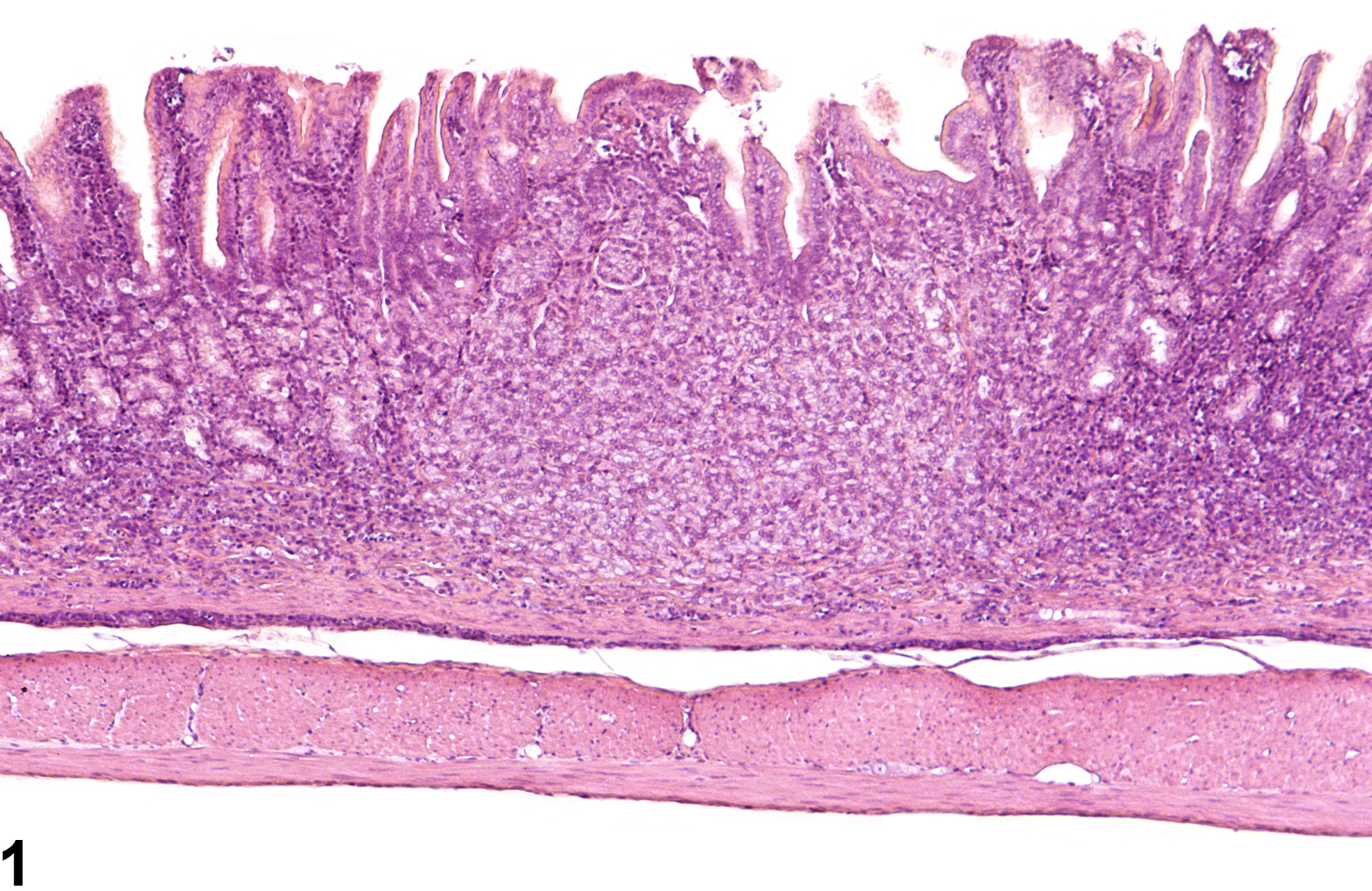
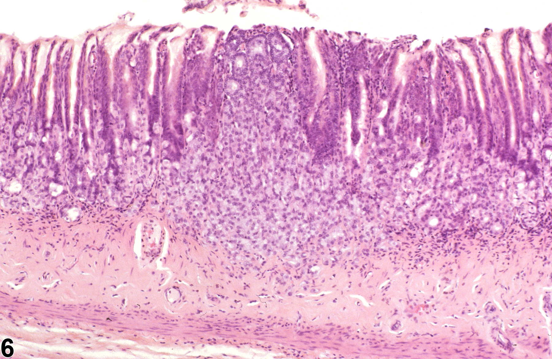
Stomach, Glandular stomach, Neuroendocrine cell - Hyperplasia in a female F344/N rat from a chronic study. The mucosa is expanded by a pale focus of cells.
All Images

Stomach, Glandular stomach, Neuroendocrine cell - Hyperplasia in a female F344/N rat from a chronic study. The mucosa is expanded by a pale focus of cells.
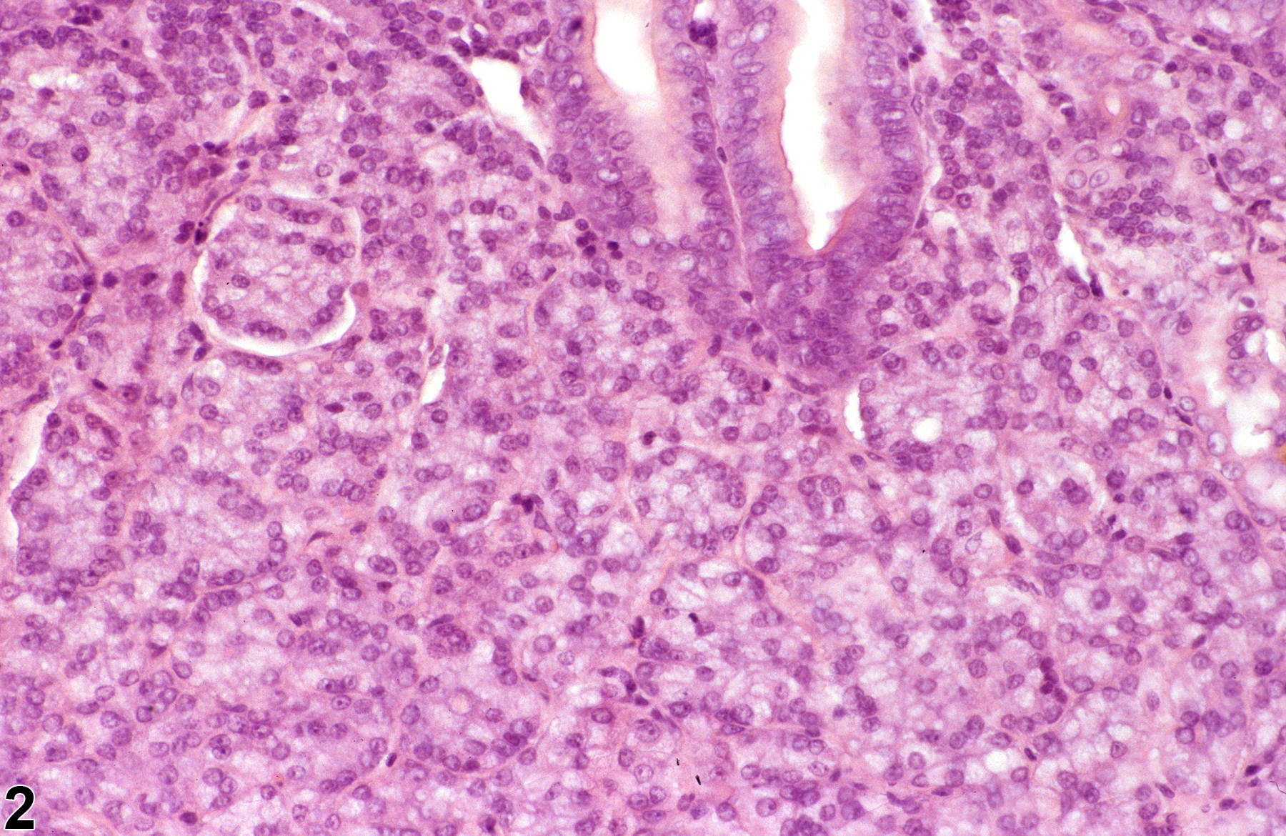
Stomach, Glandular stomach, Neuroendocrine cell - Hyperplasia in a female F344/N rat from a chronic study (higher magnification of Figure 1). The hyperplastic cells have small cytoplasmic vacuoles and are grouped in to small clusters separated by a fine vascular stroma.
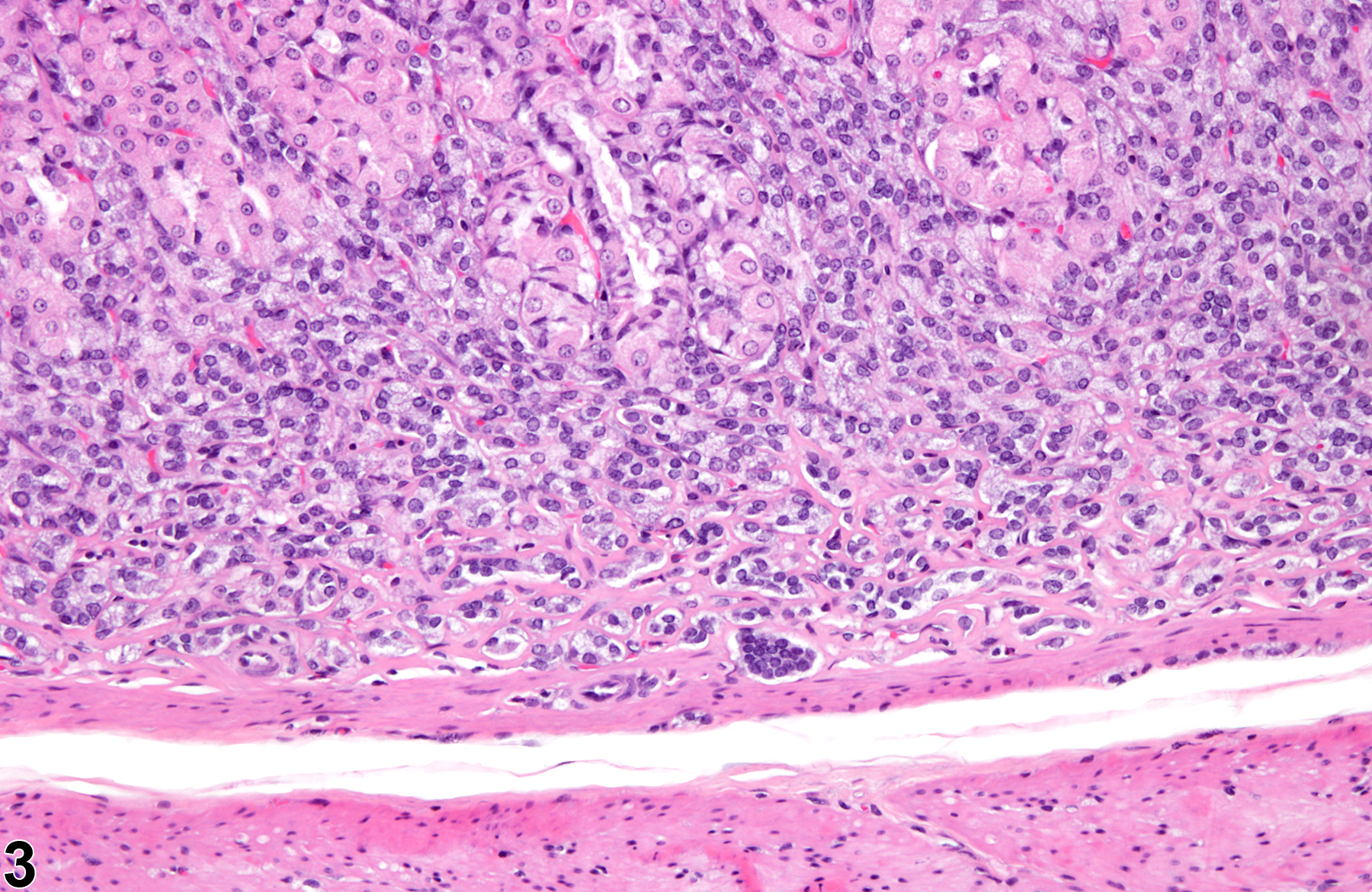
Stomach, Glandular stomach, Neuroendocrine cell - Hyperplasia in a female F344/N rat from a chronic study. The mucosal glands are separated by smaller cells with fine cytoplasmic vacuoles.
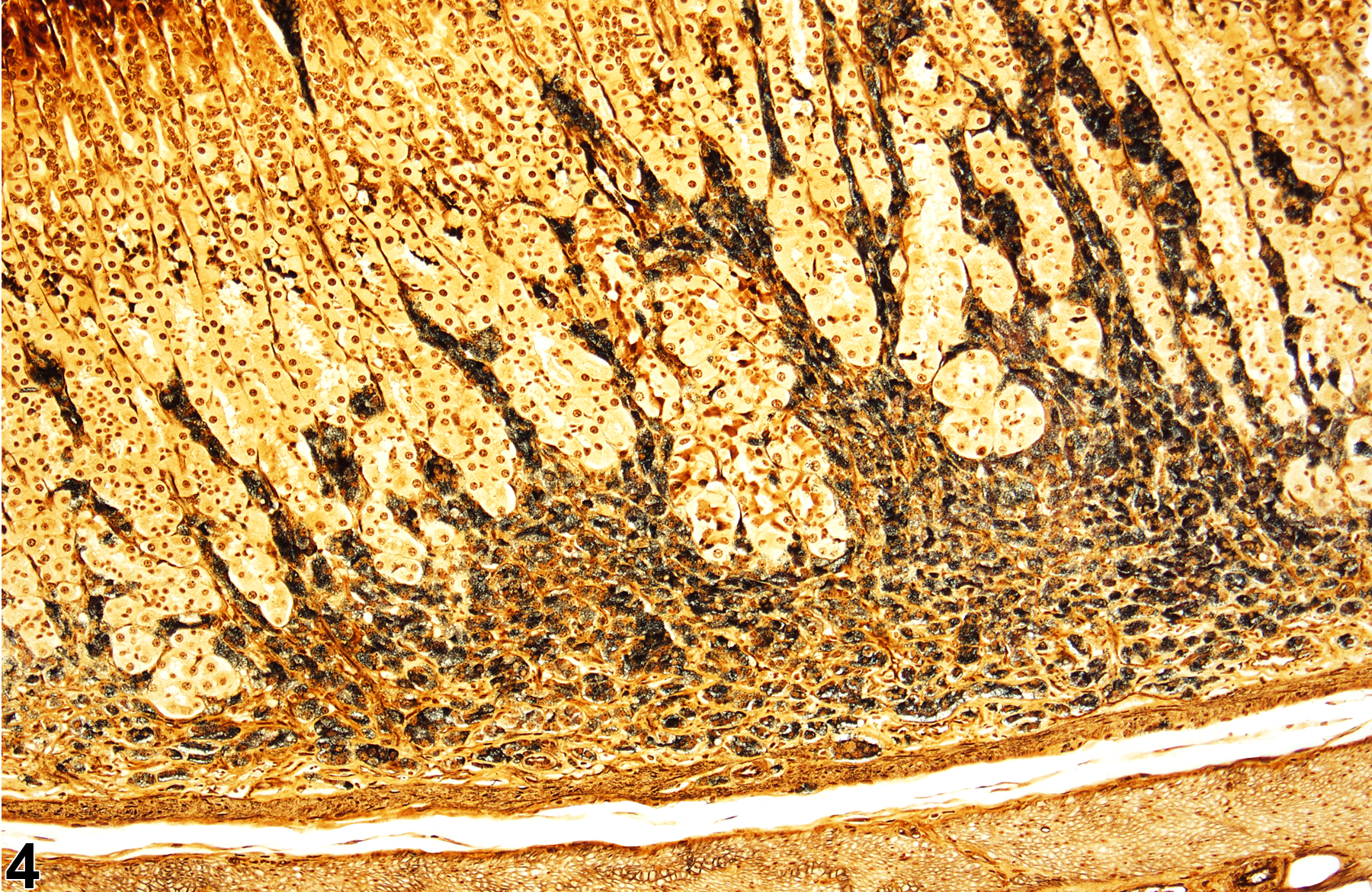
Stomach, Glandular stomach, Neuroendocrine cell - Hyperplasia in a female F344/N rat from a chronic study (Sevier-Munger stain). The cells separating the glands stain positively with a silver stain.
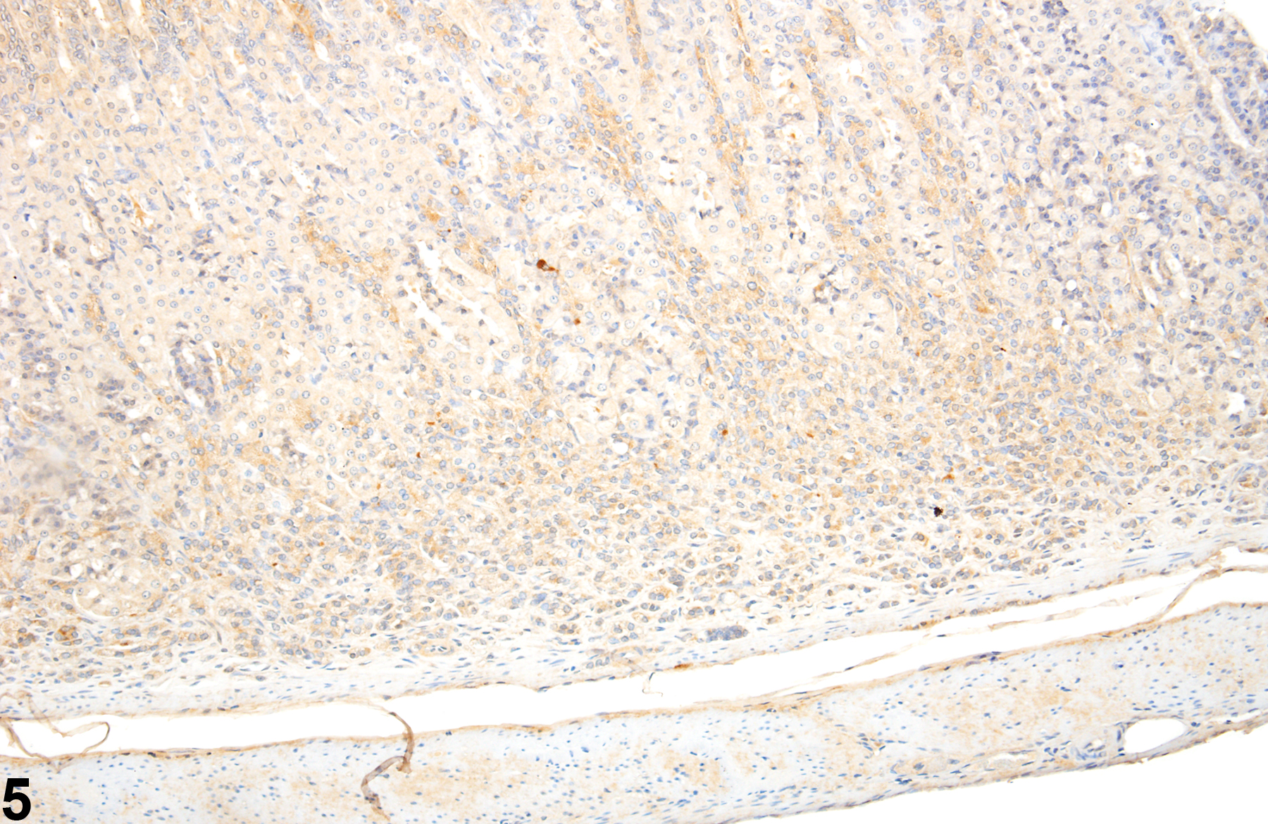
Stomach, Glandular stomach, Neuroendocrine cell - Hyperplasia in a female F344/N rat from a chronic study (Chromagranin A immunohistochemistry). The cells separating the glands are immunopositive for Chromagranin A.
Stomach, Glandular stomach, Neuroendocrine cell - Hyperplasia in a female F344/N rat from a chronic study (Sevier-Munger stain). The gastric glands are separated by pale-staining cells.