

Alimentary System
Stomach, Glandular Stomach - Pigment
Narrative
Bertram TA, Markovits JE, Juliana MM. 1996. Non-proliferative lesions of the alimentary canal in rats GI-1. In Guides for Toxicologic Pathology. STP/ARP/AFIP, Washington, DC, 1-16.
Full Text: https://www.toxpath.org/docs/SSNDC/GINonproliferativeRat.pdfBetton GR. 1998. The digestive system I: The gastrointestinal tract and exocrine pancreas. In: Target Organ Pathology (Turton J, Hooson J, eds). Taylor and Francis, London, 29-60.
Myers RK, McGavin MD. 2007. Cellular and tissue responses to injury. In: Pathologic Basis of Veterinary Disease, 4th ed (McGavin MD, Zachary JF, eds). Mosby, St Louis, MO, 3-62.
National Toxicology Program. 1992. NTP TR-409. Toxicology and Carcinogenesis Studies of Quercetin (CAS No. 117-39-5) in F344/N Rats (Feed Studies). NTP, Research Triangle Park, NC.
Abstract: https://ntp.niehs.nih.gov/go/7698Pizzolato P. 1976. Formalin pigment (acid hematin) and related pigments. Am J Med Technol 42:436-440.
Abstract: https://www.ncbi.nlm.nih.gov/pubmed/790956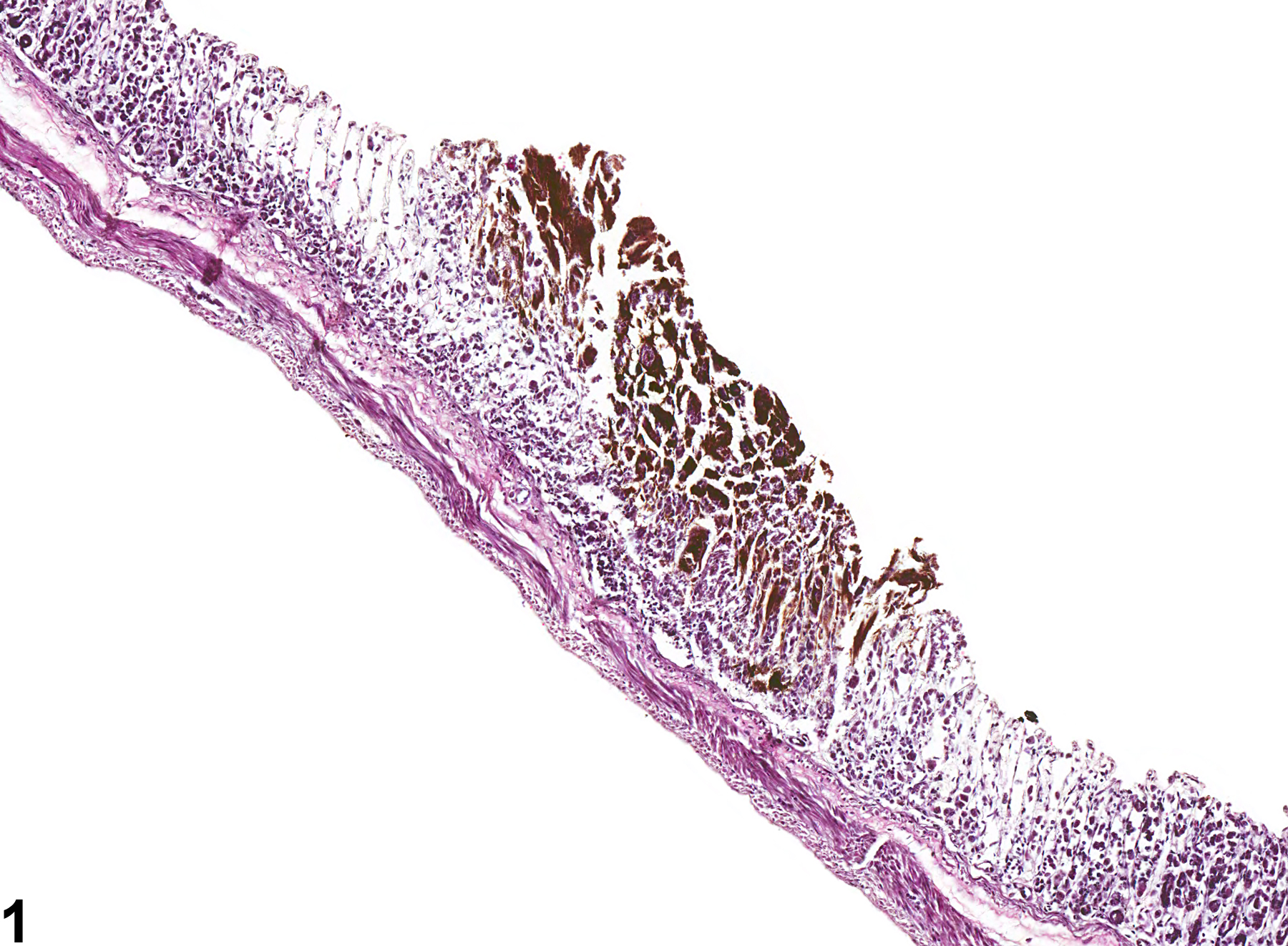
Stomach, Glandular stomach - Pigment in female B6C3F1 mouse from a chronic study. Pigment (likely hemosiderin) is present within a necrotic area of the mucosa.
All Images

Stomach, Glandular stomach - Pigment in female B6C3F1 mouse from a chronic study. Pigment (likely hemosiderin) is present within a necrotic area of the mucosa.

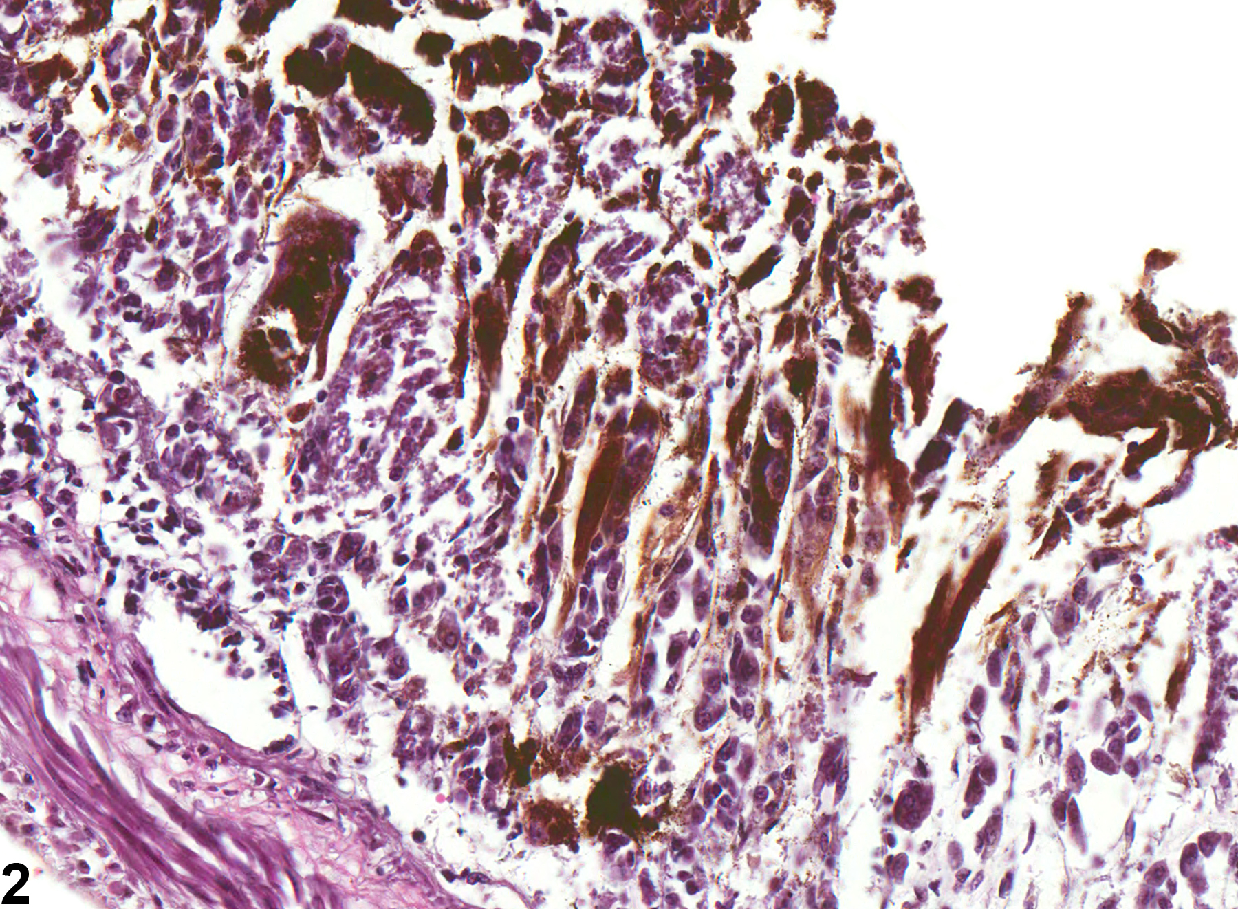
Stomach, Glandular stomach - Pigment in female B6C3F1 mouse from a chronic study (higher magnification of Figure 1). Pigment (likely hemosiderin) is present within a necrotic area of the mucosa.

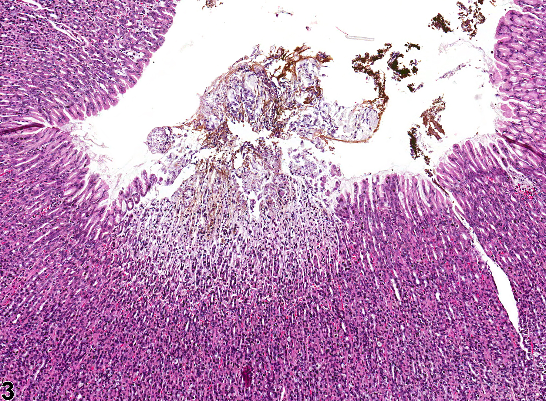
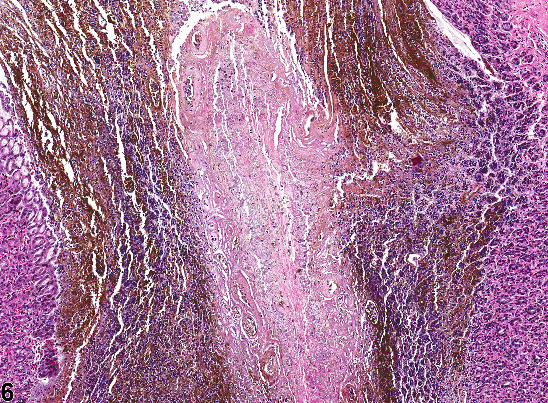
Stomach, Glandular stomach - Pigment in female F344/N rat from a chronic study. Pigment is present within a necrotic area of the mucosa.

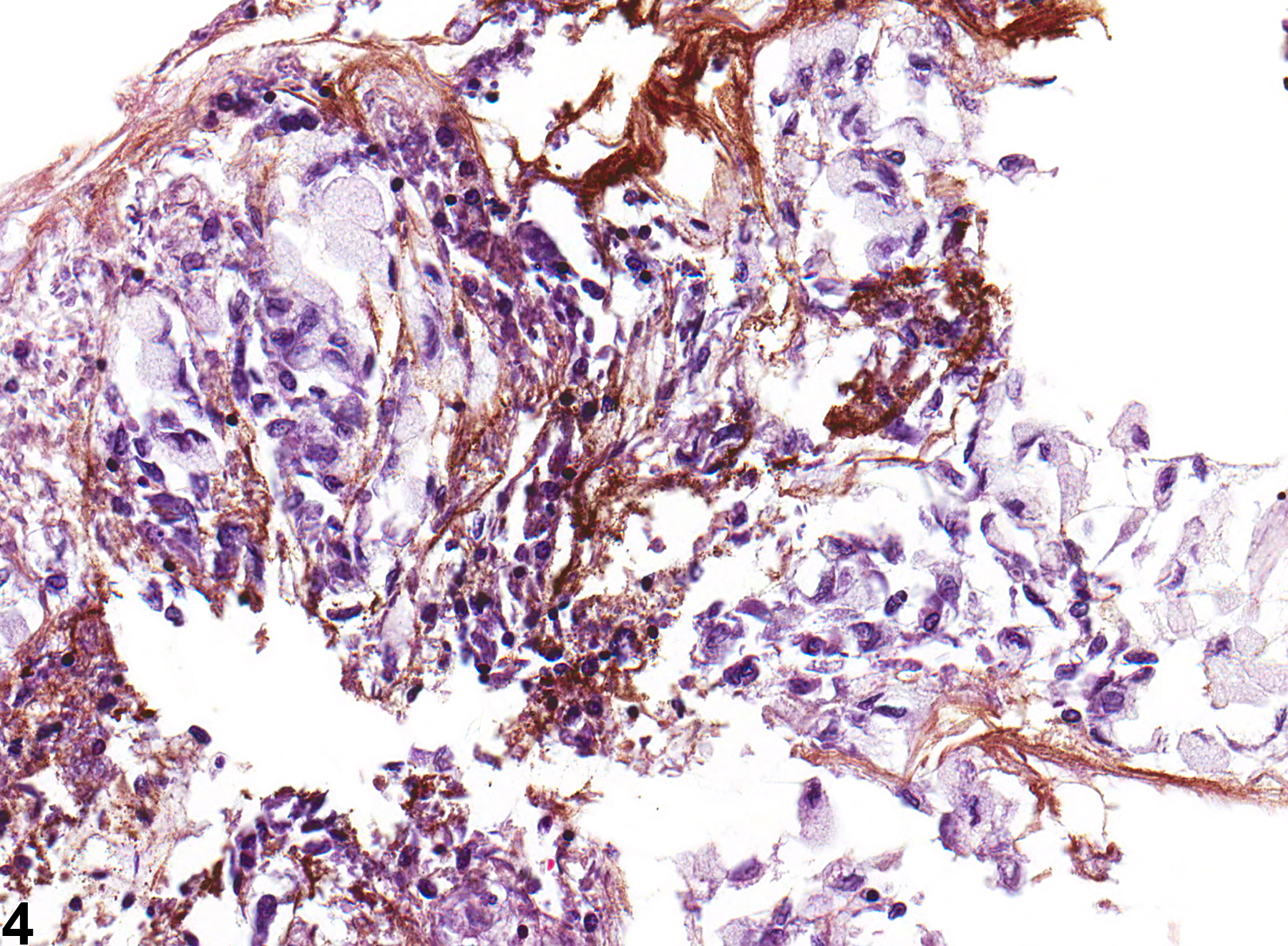
Stomach, Glandular stomach - Pigment in female F344/N rat from a chronic study (higher magnification of Figure 1). Pigment is present within a necrotic area of the mucosa.

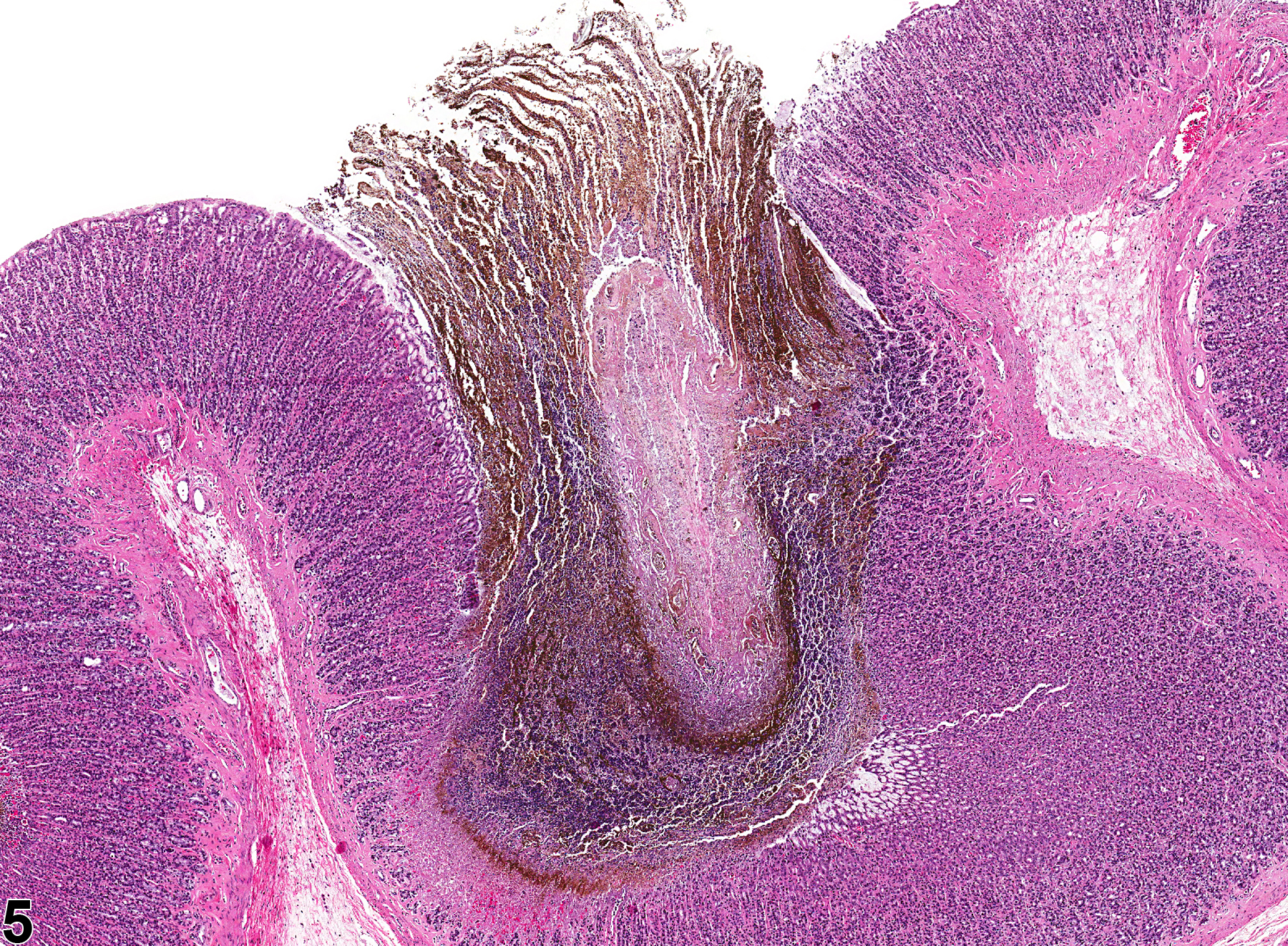
Stomach, Glandular stomach - Pigment in female F344/N rat from a chronic study. Pigment (likely hemosiderin) is present within the mucosa.

Stomach, Glandular stomach - Pigment in female F344/N rat from a chronic study (higher magnification of Figure 1). Pigment (likely hemosiderin) is present within the mucosa.