

Alimentary System
Stomach, Glandular Stomach, Epithelium - Vacuolation, Cytoplasmic
Narrative
Haggerty HG, Warner WA, Comereski CR, Peden WM, Mezza LE, Damle BD, Siegall CB, Davidson TJ. 1999. BR96 sFv-PE40 immunotoxin: Nonclinical safety assessment. Toxicol Pathol 27:87-94.
Full Text: http://tpx.sagepub.com/content/27/1/87.full.pdfMyers RK, McGavin MD. 2007. Cellular and tissue responses to injury. In: Pathologic Basis of Veterinary Disease, 4th ed (McGavin MD, Zachary JF, eds). Mosby, St Louis, MO, 3-62.
National Toxicology Program. 2007. NTP TOX-72. Toxicity Studies of Sodium Dichromate Dihydrate (CAS No. 7789-12-0) Administered in Drinking Water to F344/N Rats and B6C3F1 Mice and Male Balb/c and am3-C57BL/6 Mice. NTP, Research Triangle Park, NC.
Abstract: https://ntp.niehs.nih.gov/go/11170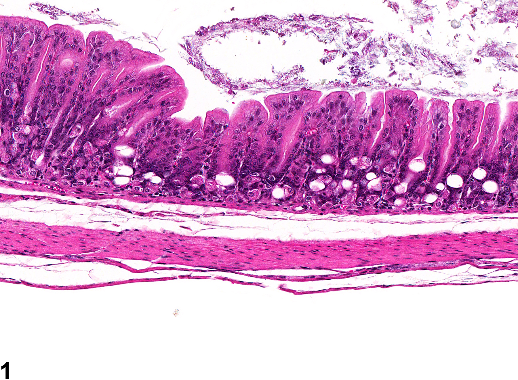
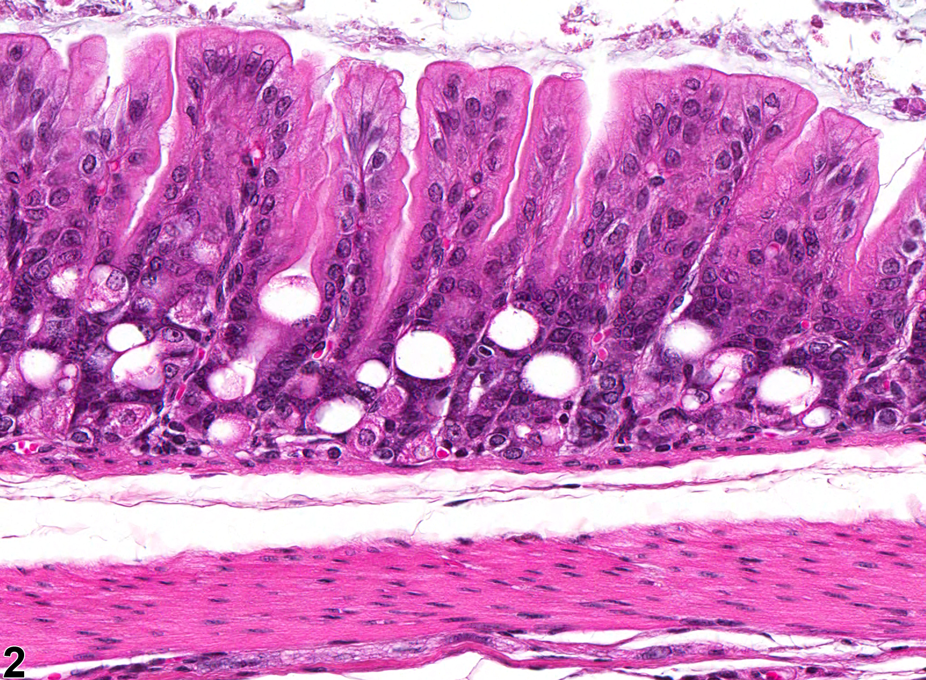
Stomach, Glandular stomach, Epithelium - Vacuolation, Cytoplasmic in a male F344/N rat from a subchronic study. There is vacuolation of epithelial cells in the mucosal glands.
All Images

Stomach, Glandular stomach, Epithelium - Vacuolation, Cytoplasmic in a male F344/N rat from a subchronic study. There is vacuolation of epithelial cells in the mucosal glands.

Stomach, Glandular stomach, Epithelium - Vacuolation, Cytoplasmic in a male F344/N rat from a subchronic study (higher magnification of Figure 1). There is vacuolation of epithelial cells in the mucosal glands.