

Alimentary System
Oral Mucosa - Foreign Body
Narrative
Bertram TA, Markovits JE, Juliana MM. 1996. Non-proliferative lesions of the alimentary canal in rats GI-1. In Guides for Toxicologic Pathology. STP/ARP/AFIP, Washington, DC, 1-16.
Full Text: https://www.toxpath.org/docs/SSNDC/GINonproliferativeRat.pdfBrown HR, Hardisty JF. 1990. Oral cavity, esophagus and stomach. In: Pathology of the Fischer Rat (Boorman GA, Montgomery CA, MacKenzie WF, eds). Academic Press, San Diego, CA, 9-30.
Abstract: https://www.ncbi.nlm.nih.gov/nlmcatalog/9002563Madsen C. 1989. Squamous-cell carcinoma and oral, pharyngeal and nasal lesions caused by foreign bodies in feed. Cases from a long-term study in rats. Lab Anim 23:241-247.
Abstract: https://www.ncbi.nlm.nih.gov/pubmed/2668638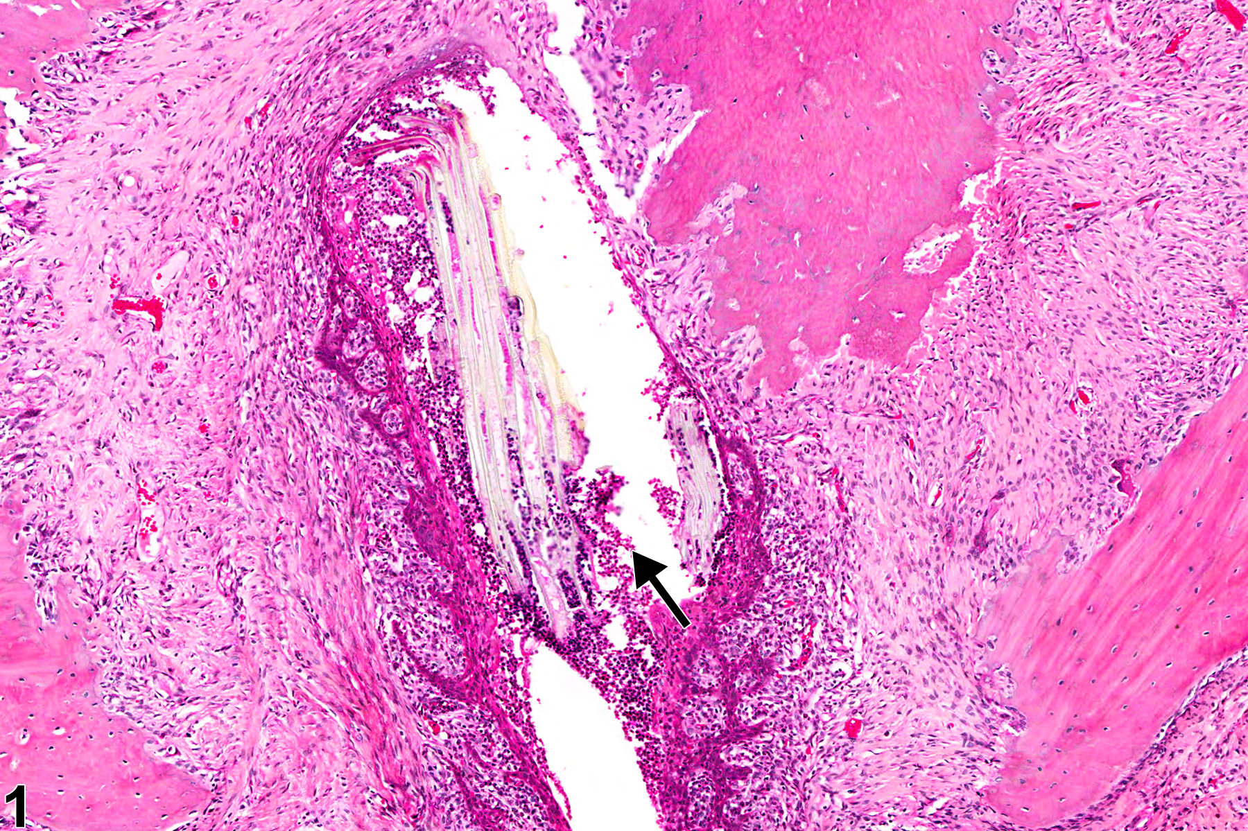
Oral mucosa - Foreign body in a male F344/N rat from a chronic study. The foreign body (arrow) is surrounded by suppurative inflammation.
All Images

Oral mucosa - Foreign body in a male F344/N rat from a chronic study. The foreign body (arrow) is surrounded by suppurative inflammation.

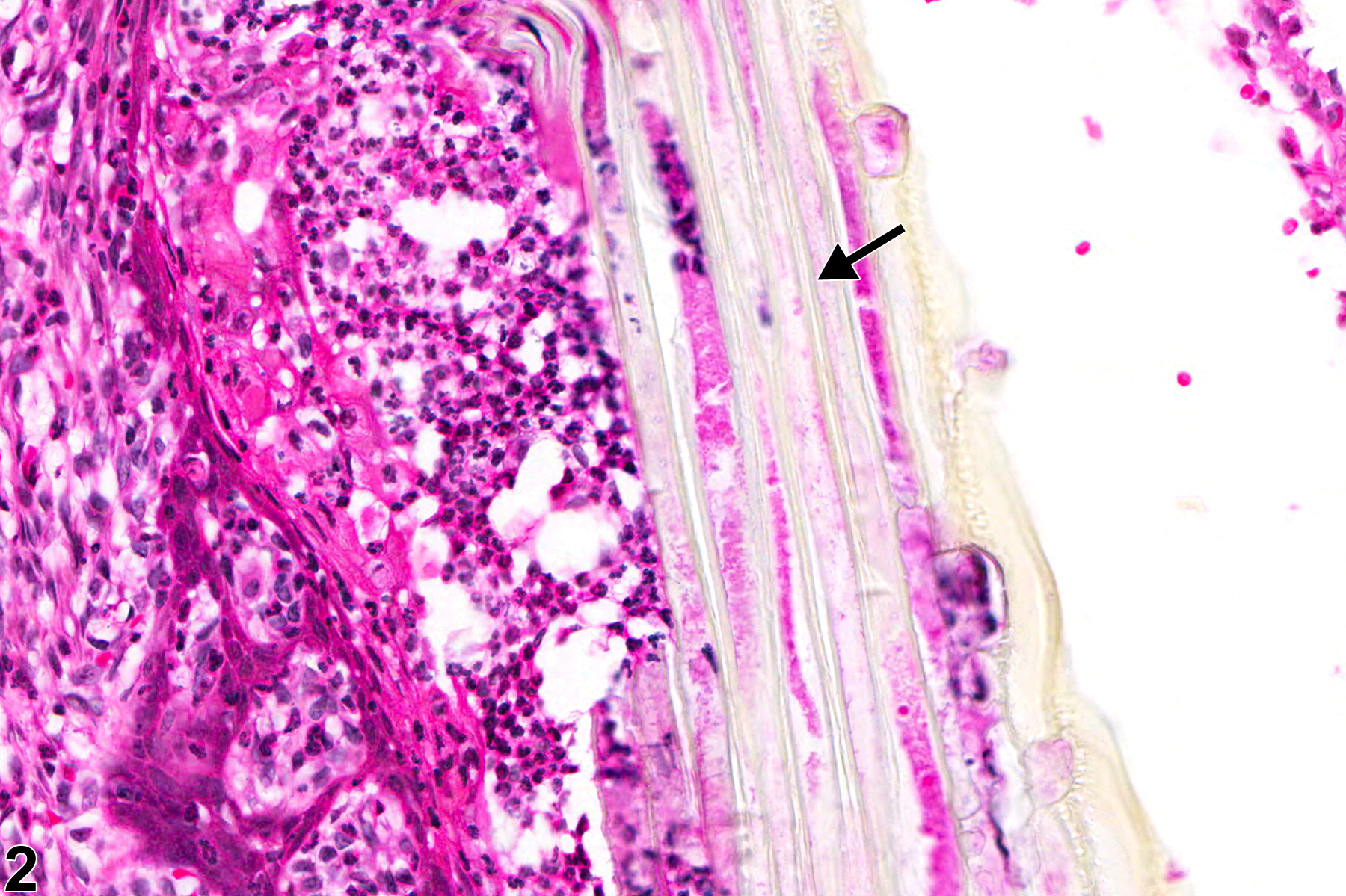
Oral mucosa - Foreign body in a male F344/N rat from a chronic study (higher magnification of Figure 1). The foreign body (arrow) appears to be plant material.

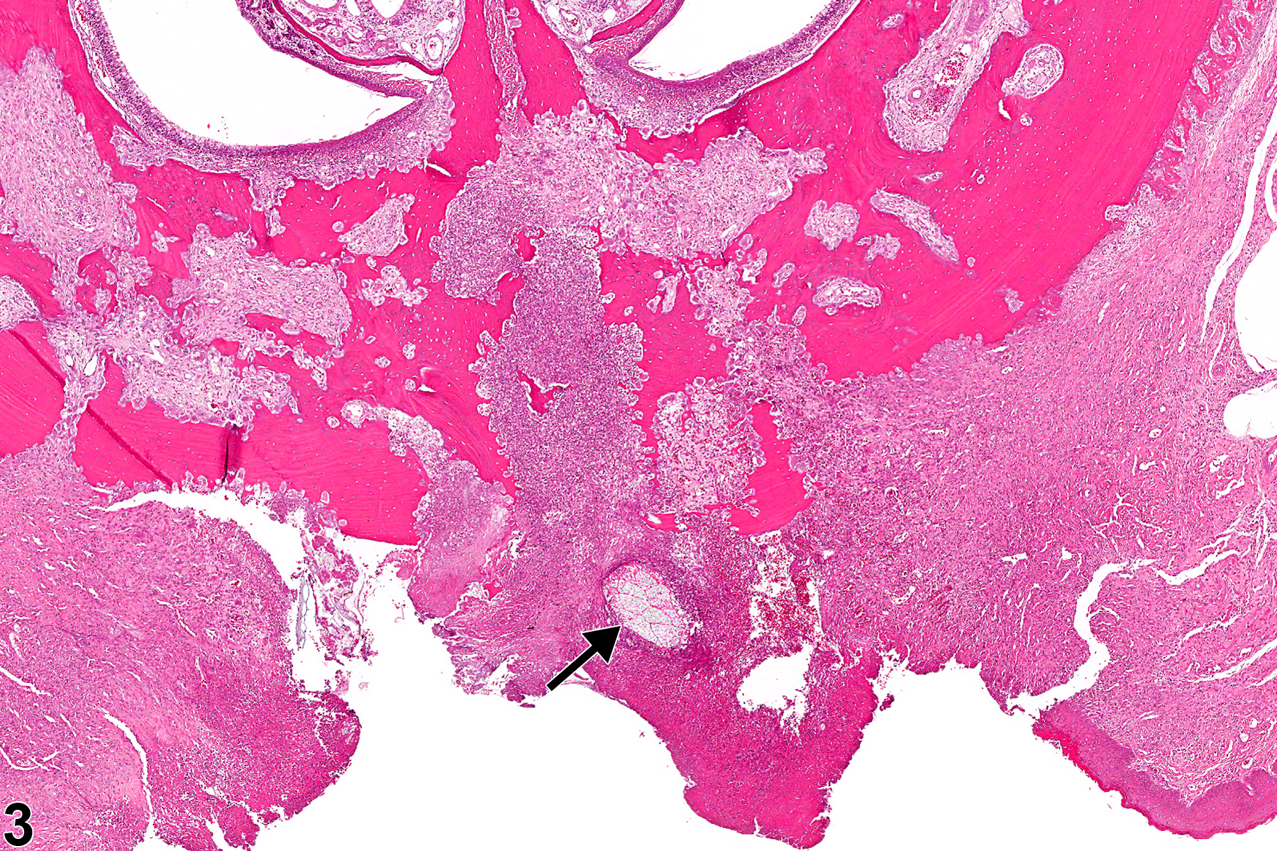
Oral mucosa - Foreign body in a male F344/N rat from a chronic study. A foreign body (arrow) associated with an ulcer in the hard palate.

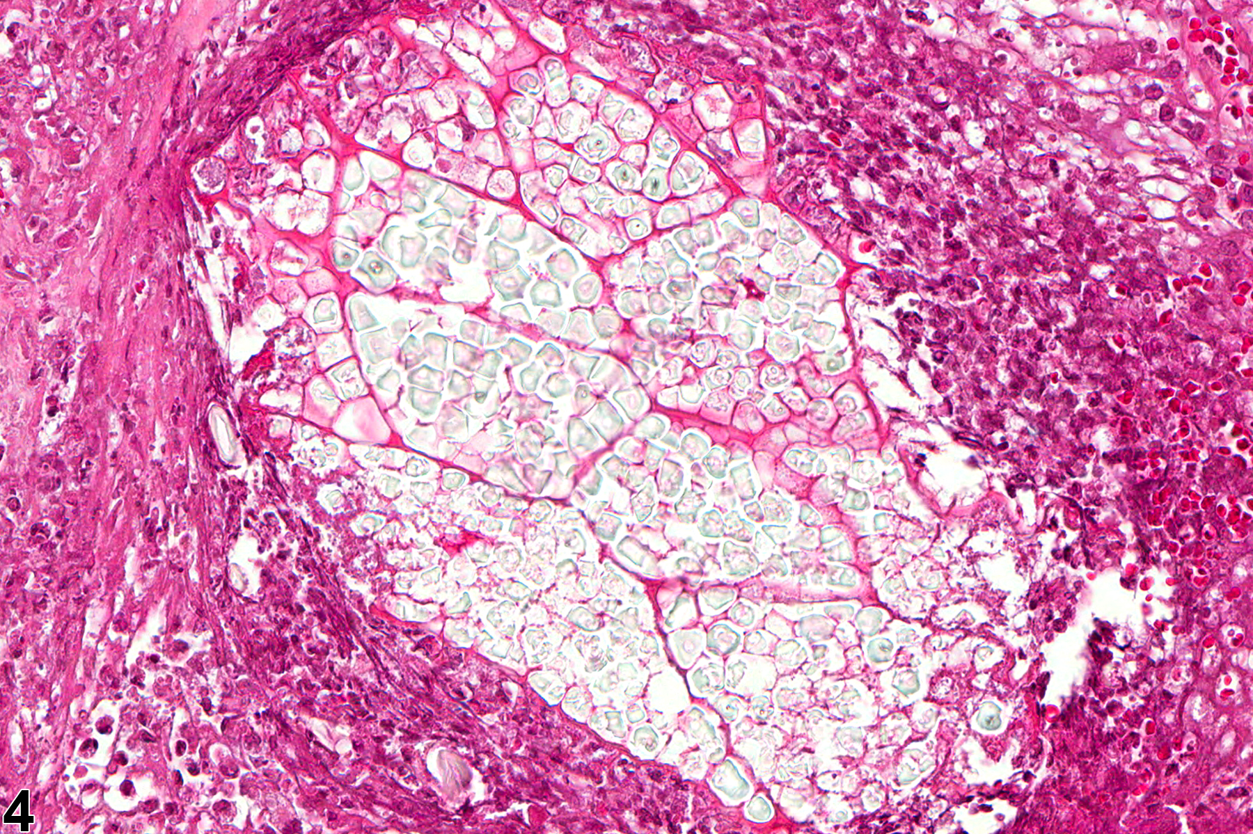
Oral mucosa - Foreign body in a male F344/N rat from a chronic study (higher magnification of Figure 3). The foreign body is surrounded by suppurative inflammation.

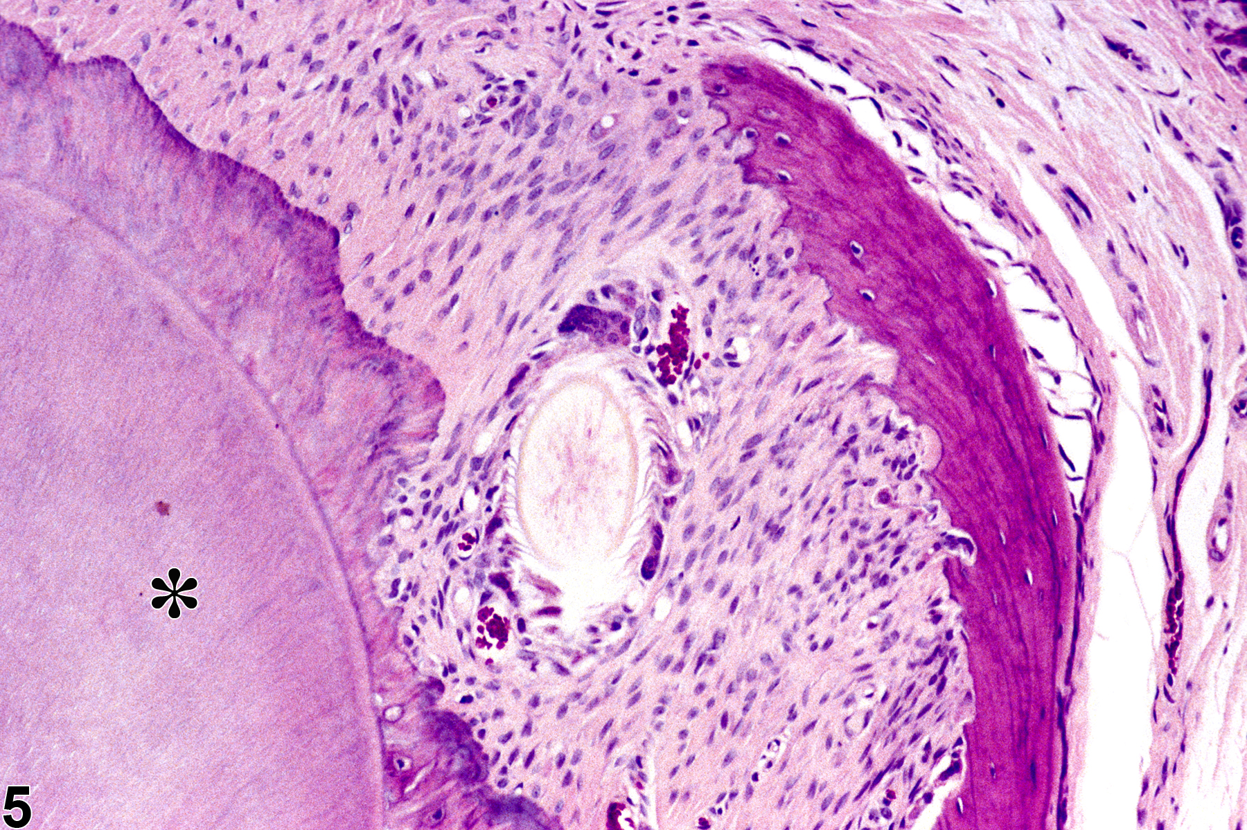
Oral mucosa - Foreign body in a female F344/N rat from a chronic study. There is a foreign body adjacent to a tooth (asterisk).


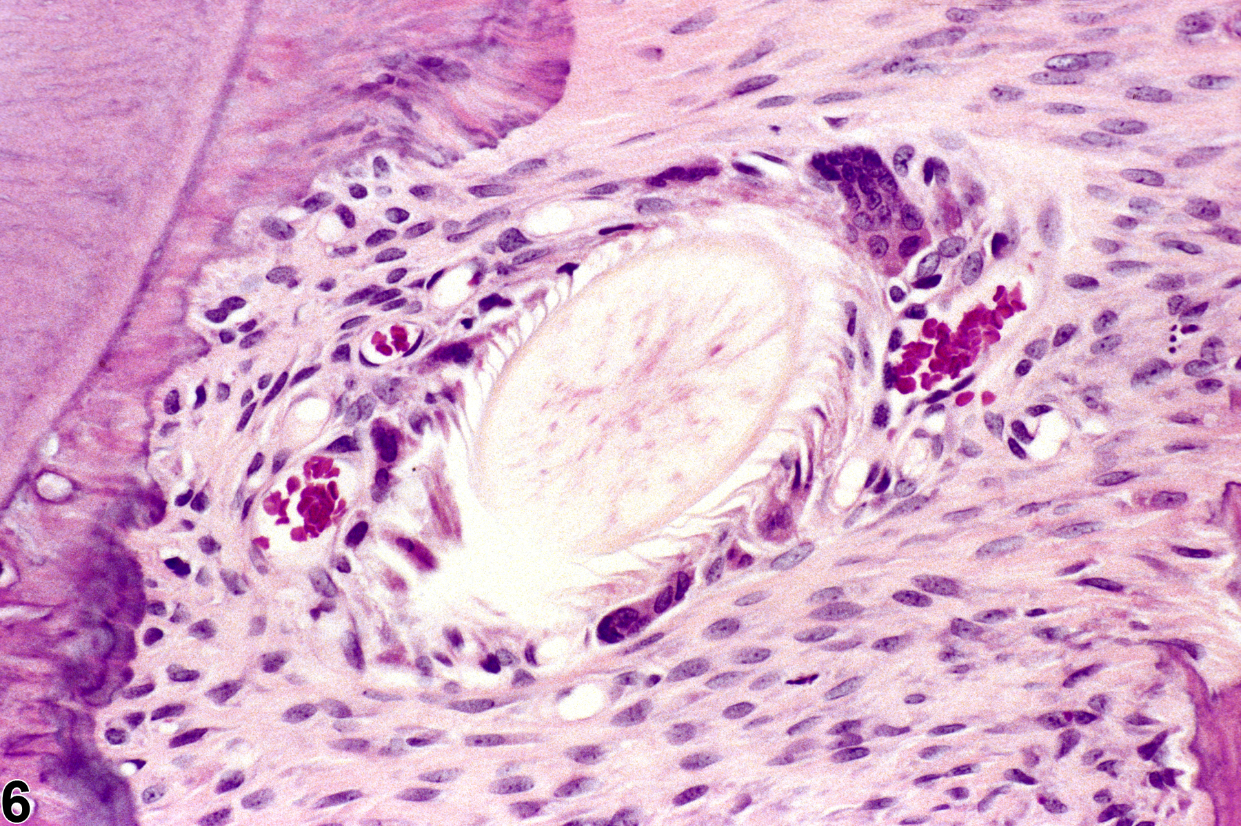
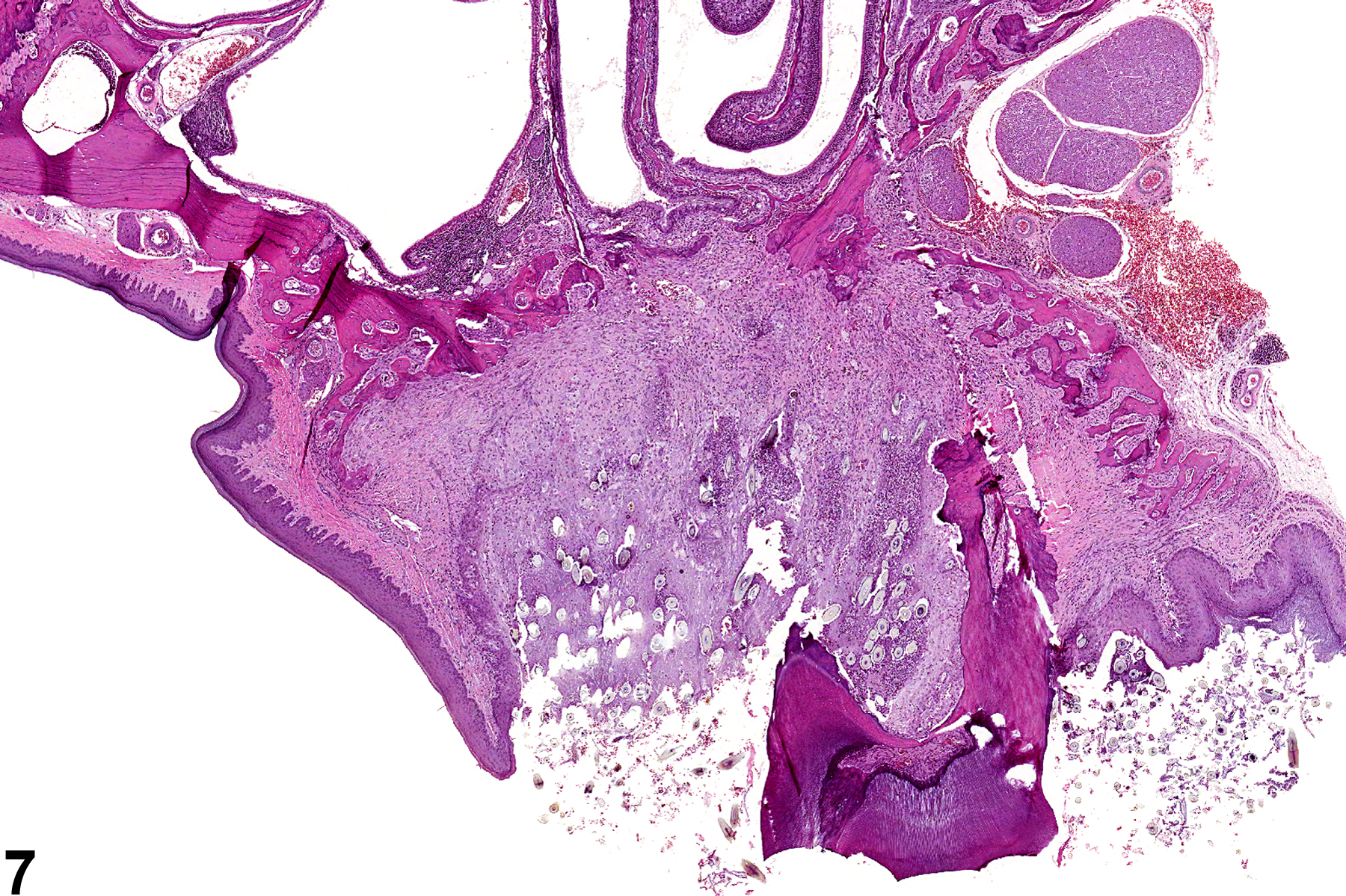
Oral mucosa - Foreign body in a male B6C3F1 mouse from a chronic study. Multiple hair shafts and associated inflammation are present in the periodontal tissue.

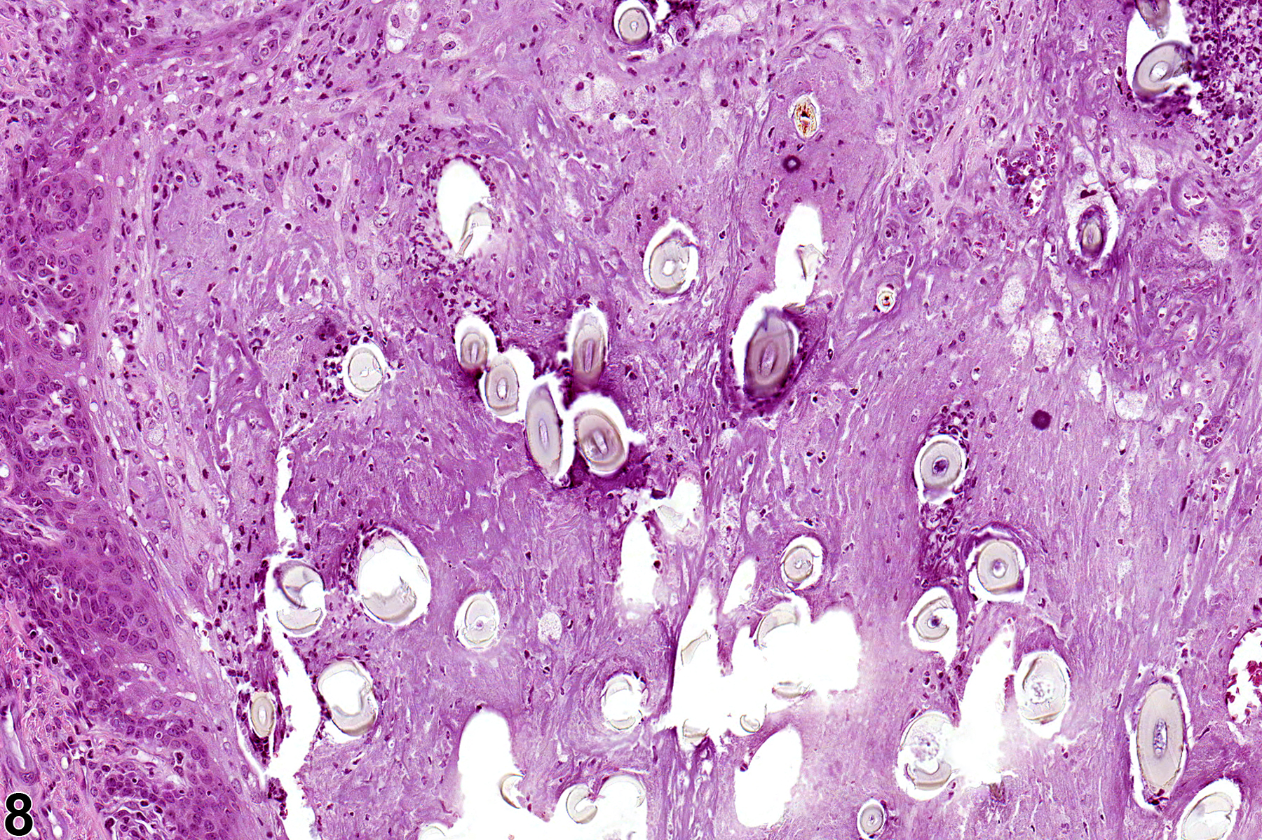
Oral mucosa - Foreign body in a male B6C3F1 mouse from a chronic study (higher magnification of Figure 7). Multiple hair shafts and associated inflammation are present in the periodontal tissue.