U.S. flag

An official website of the United States government

Dot gov

The .gov means it's official.
Federal government websites often end in .gov or .mil. Before sharing sensitive information, make sure you're on a federal government site.

Https

The site is secure.
The https:// ensures that you are connecting to the official website and that any information you provide is encrypted and transmitted securely.

Skip to Main Navigation
Skip to Page Content
Skip to Atlas Navigation

Salivary Gland - Amyloid

Image of amyloid in the salivary gland from a female Swiss Webster mouse in a chronic study
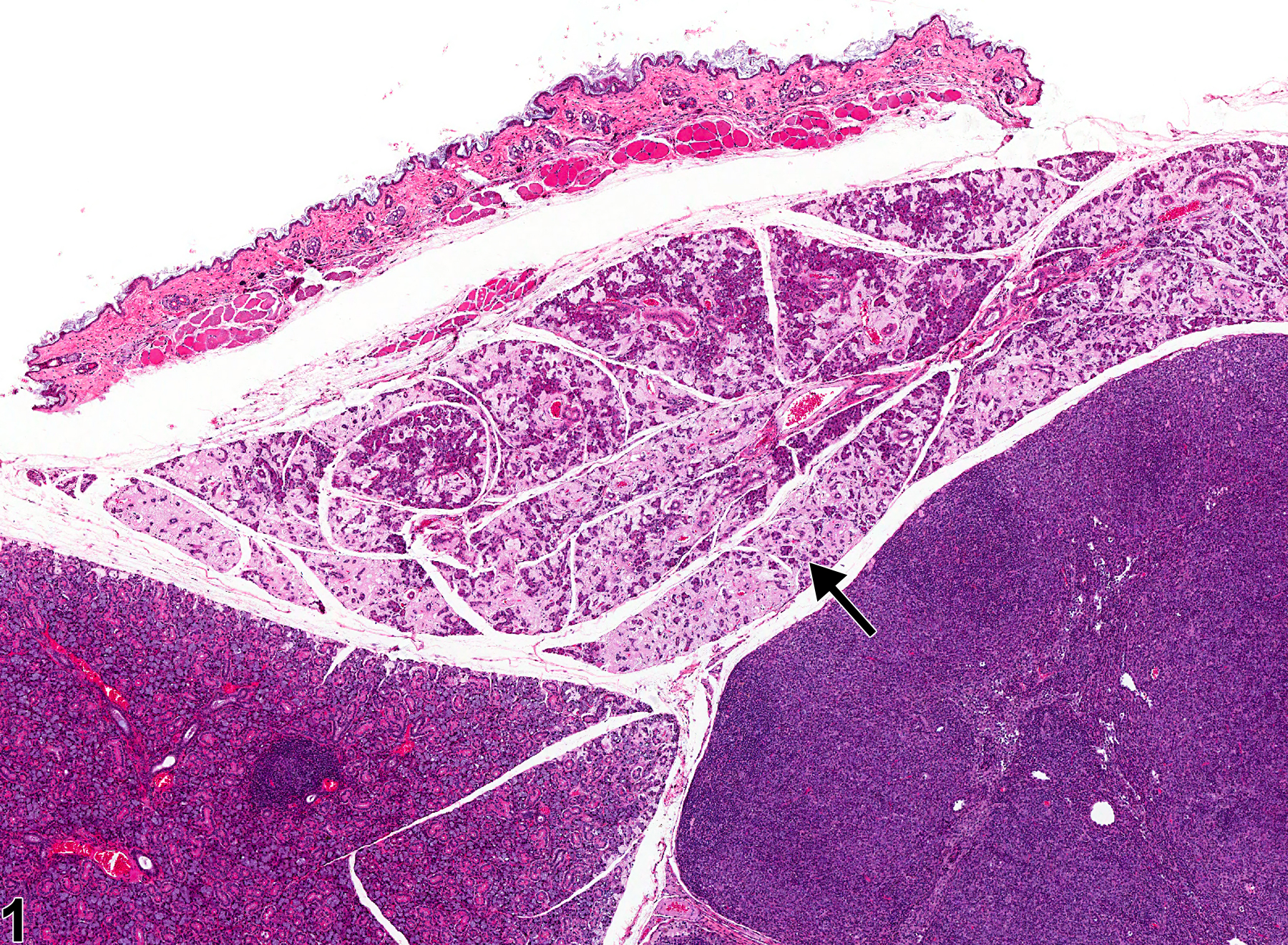
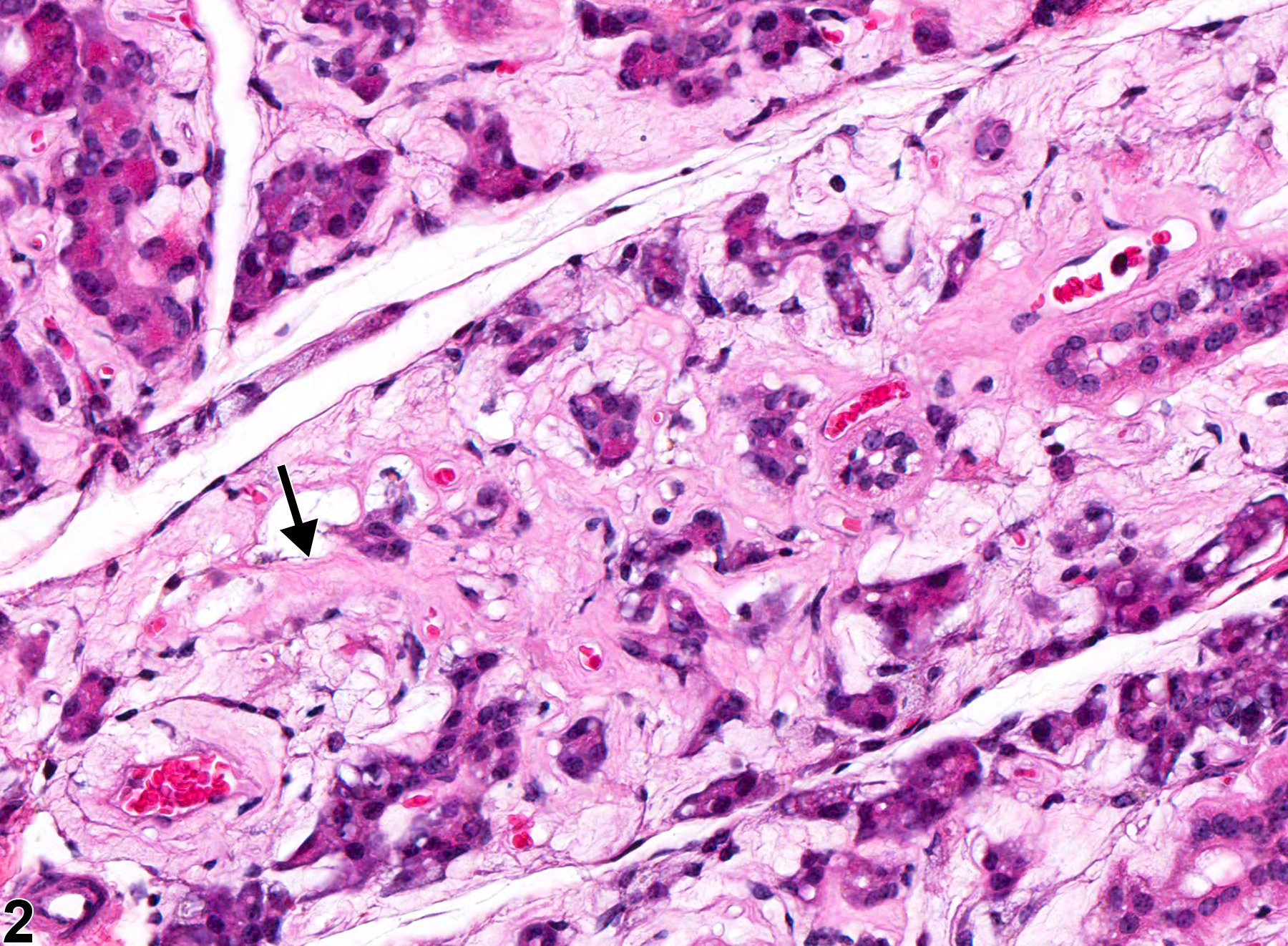
Salivary gland - Amyloid in a female Swiss Webster mouse from a chronic study. The deposition of amyloid is affecting an entire lobe of the salivary gland (arrow).
Figure 1 of 2
Image of amyloid in the salivary gland from a female Swiss Webster mouse in a chronic study
Salivary gland - Amyloid in a female Swiss Webster mouse from a chronic study (higher magnification of Figure 1). The extracellular amyloid (arrow) is causing atrophy of the glandular acini.
Figure 2 of 2
next arrow

comment:

Amyloid deposition in tissues (amyloidosis) is a systemic disease that is rare in B6C3F1, BALB/c, and C3H mice but common in CD-1, A, Swiss Webster, SJL, and B6 mice and can be a cause of death. The incidence can be increased in association with fighting among group-housed males and with ectoparasitism. Amyloid appears as an amorphous, eosinophilic, hyaline, extracellular substance that, with progressive accumulation, results in atrophy of adjacent cells and tissue. In the salivary gland, the amyloid can cause atrophy of the salivary gland acini ( Figure 1image opens in a pop-up window and Figure 2image opens in a pop-up window ). In the esophagus and small intestine, the disease appears to begin as a deposition of amyloid in the submucosa. Amyloid deposits in the glomeruli of the kidney are usually the cause of death in animals that die with amyloidosis. Congo red stains amyloid orange to orange red and under polarized light imparts a light green, so-called apple green, fluorescence.

recommendation:

Whenever present, amyloid deposits in the salivary gland should be diagnosed as "amyloid" and graded based on the extent of the amyloid deposits. Associated atrophy of the tissues need not be diagnosed separately but can be described in the narrative.

references:

Gad S. 2007. The mouse. In: Animal Models of Toxicology, 2nd ed (Gad S, ed). CRC Press, Boca Raton, FL, 19-146.
Abstract: http://www.crcnetbase.com/isbn/9781420014204

Myers RK, McGavin MD. 2007. Cellular and tissue responses to injury. In: Pathologic Basis of Veterinary Disease, 4th ed (McGavin MD, Zachary JF, eds). Mosby, St Louis, MO, 14-62.

National Toxicology Program. 1993. NTP TR-443. Toxicology and Carcinogenesis Studies of Oxazepam (CAS No. 604-75-1) in Swiss-Webster and B6C3F1 Mice (Feed Studies). NTP, Research Triangle Park, NC.
Abstract: https://ntp.niehs.nih.gov/go/6030

Percy DH, Barthold SW. 2001. Mouse. In: Pathology of Laboratory Rodents and Rabbits, 2nd ed. Iowa State Press, Ames, 2001, 3-106.
Abstract: http://onlinelibrary.wiley.com/book/10.1002/9780470344613