

Alimentary System
Salivary Gland - Atrophy
Narrative
Decreased food consumption or protein starvation can reduce the weight of salivary glands in rats. Shrinking of mucous and serous glands and loss of zymogen granules are associated with decreased RNA but unchanged DNA content, attributable to the reduced requirements for protein synthesis. As salivary gland function is responsive to adrenergic stimulation, it is not surprising that atrophy occurs following adrenergic blockage. The weights of the submandibular gland in mice were shown to decrease after administration of the B-adrenergic blocking agent propranolol. This was associated with a reduction in stainable neutral mucins and a decrease in the thickness of the acinar cells, making the gland lumens appear larger than normal.
Salivary gland atrophy occurs occasionally in aging B6C3F1 mice. The atrophy generally affects individual lobules and is characterized by a decrease in the size of acini and acinar epithelial cells, which may be accompanied by an infiltrate of chronic inflammatory cells and an increase in interstitial connective tissue and/or in the number of glandular ducts. The submandibular and parotid glands tend to be involved more commonly than the sublingual gland.
Botts S, Jokinen M, Gaillard ET, Elwell MR, Mann PC. 1999. Salivary, Harderian, and lacrimal glands. In: Pathology of the Mouse (Maronpot RR, ed). Cache River Press, St Louis, MO, 49-80.
Boyd EM, Cehn CP, Muis LF. 1970. Resistance to starvation in albino rats fed from weaning on diets containing from 0 to 81% of protein as casein. Growth 23:99-112.
Greaves P. 2007. Digestive system. In: Histopathology of Preclinical Toxicity Studies, 3rd ed. Academic Press, London, 334-456.
McBride, Harper RK, Siegel IA. 1987. Methotrexate-induced changes in rat parotid and submandibular gland function. J Dental Res 66: 1445-1448.
Abstract: https://www.ncbi.nlm.nih.gov/pubmed/2442228Neuenschwander SB, Elwell MR. 1990. Salivary glands. In: Pathology of the Fischer Rat (Boorman GA, Montgomery CA, MacKenzie WF, eds). Academic Press, San Diego, CA, 31-42.
Smith B, Butler M. 1978. The effects of long-term propranolol on the salivary glands and intestinal mucosa of the mouse. J Pathol 124:185-187.
Abstract: https://www.ncbi.nlm.nih.gov/pubmed/722381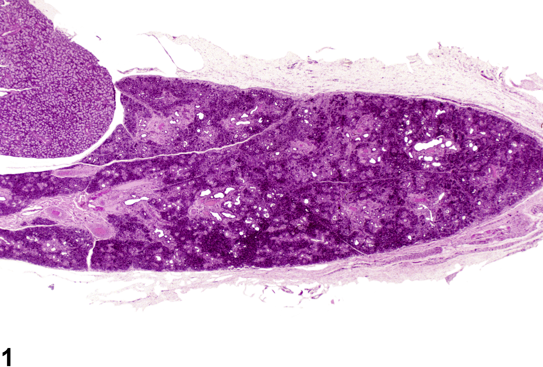
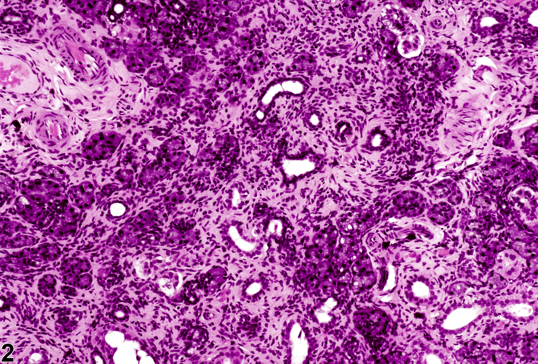
Salivary gland - Atrophy in a male F344/N rat from a chronic study. There is a decrease in the number and size of the glands and an increase in the connective tissue.
All Images

Salivary gland - Atrophy in a male F344/N rat from a chronic study. There is a decrease in the number and size of the glands and an increase in the connective tissue.

Salivary gland - Atrophy in a male F344/N rat from a chronic study (higher magnification of Figure 1). There is a decrease in the number and size of the glands and an increase in the connective tissue.