U.S. flag

An official website of the United States government

Dot gov

The .gov means it's official.
Federal government websites often end in .gov or .mil. Before sharing sensitive information, make sure you're on a federal government site.

Https

The site is secure.
The https:// ensures that you are connecting to the official website and that any information you provide is encrypted and transmitted securely.

Skip to Main Navigation
Skip to Page Content
Skip to Atlas Navigation

Salivary Gland - Fibrosis

Image of fibrosis in the salivary gland from a male F344/N rat in a subchronic study
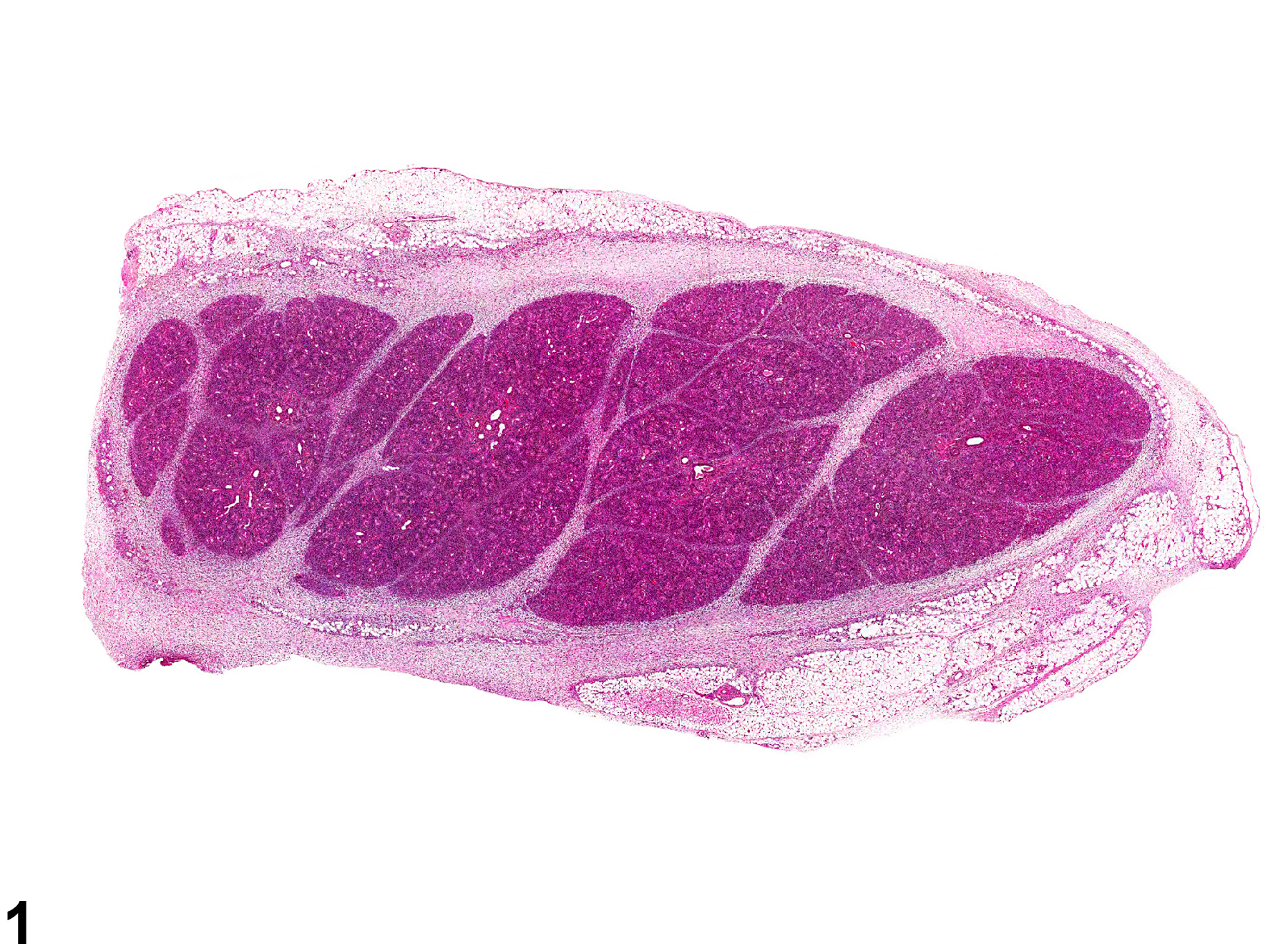
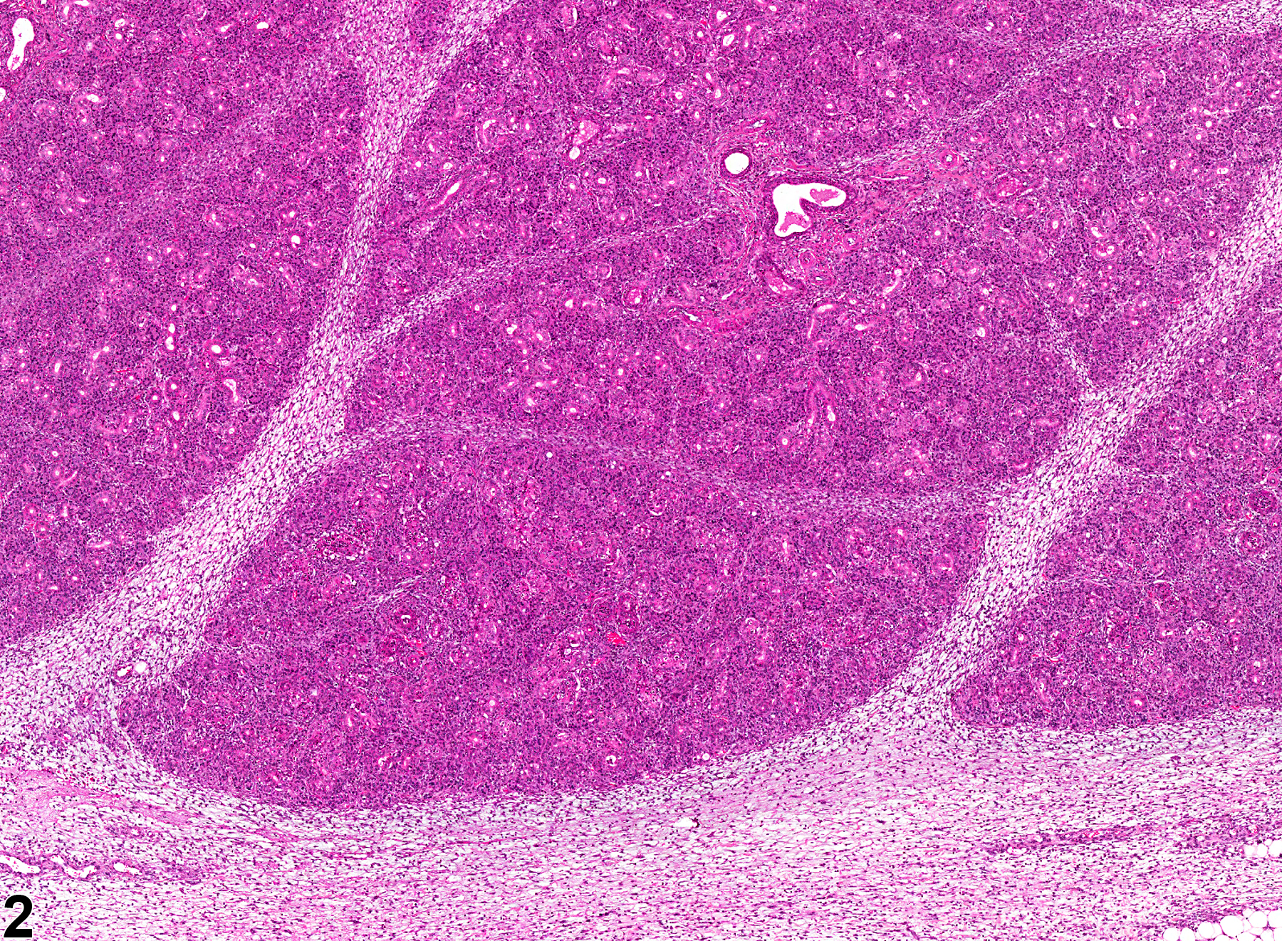
Salivary gland - Fibrosis in a male F344/N rat from a subchronic study. Fibrillar eosinophilic material (fibrosis) surrounds the salivary gland and separates the lobules.
Figure 1 of 2
Image of fibrosis in the salivary gland from a male F344/N rat in a subchronic study
Salivary gland - Fibrosis in a male F344/N rat from a subchronic study (higher magnification of Figure 1). There are inflammatory cells within the fibrotic tissue.
Figure 2 of 2
next arrow

comment:

Fibrosis can be found in and around the salivary glands ( Figure 1image opens in a pop-up window and Figure 2image opens in a pop-up window ) and often replaces glandular tissue. It may be seen concurrently with severe inflammation and necrosis. In some chronic cases, the inflammation and necrosis have resolved and the fibrosis appears as largely acellular, eosinophilic tissue. In such cases, the affected gland or lobule may be small, and there may be atrophy of the glandular acini.

recommendation:

Whenever present as the sole lesion, fibrosis should be diagnosed and graded. However, unless it is the end stage of a lesion, fibrosis is rarely seen without concurrent tissue damage or inflammation. Whenever it is a significant component of a degenerative, necrotic, or inflammatory lesion, fibrosis should be diagnosed and graded. Atrophy of glandular tissue that is considered secondary to fibrosis should not be diagnosed separately unless it is a prominent component of the lesion. Secondary lesions should be described in the pathology narrative.

references:

Ackermann MR. 2007. Chronic inflammation and wound healing. In: Pathologic Basis of Veterinary Disease, 4th ed (McGavin MD, Zachary JF, eds). Mosby, St Louis, MO, 153-191.

Brown HR, Hardisty JF. 1990. Oral cavity, esophagus and stomach. In: Pathology of the Fischer Rat (Boorman GA, Montgomery CA, MacKenzie WF, eds). Academic Press, San Diego, CA, 9-30.
Abstract: https://www.ncbi.nlm.nih.gov/nlmcatalog/9002563

Elmore S, Lanning L, Allison N, Vallant M, Nyska A. 2006. The transduction of rat submandibular glands by an adenoviral vector carrying the human growth hormone gene is associated with limited and reversible changes at the infusion site. Toxicol Pathol 34:385-392.
Abstract: https://www.ncbi.nlm.nih.gov/pubmed/16844666