

Alimentary System
Salivary Gland - Hyperplasia
Narrative
Carthew P, Slinger RP. 1981. Diagnosis of sialodacryoadenitis virus infection of rats in a virulent enzootic outbreak. Lab Anim 15:339-342.
Abstract: https://www.ncbi.nlm.nih.gov/pubmed/6281552Cotroneo E, Proctor GB, Carpenter GH. 2010. Regeneration of acinar cells following ligation of rat submandibular gland retraces the embryonic-perinatal pathway of cytodifferentiation. Differentiation 79:120-130.
Abstract: https://www.ncbi.nlm.nih.gov/pubmed/20056310Cotroneo E, Proctor GB, Paterson KL, Carpenter GH. 2008. Early markers of regeneration following ductal ligation in rat submandibular gland. Cell Tissue Res 332:227-235.
Abstract: https://www.ncbi.nlm.nih.gov/pubmed/18335244Elmore S, Lanning L, Allison N, Vallant M, Nyska A. 2006. The transduction of rat submandibular glands by an adenoviral vector carrying the human growth hormone gene is associated with limited and reversible changes at the infusion site. Toxicol Pathol 34:385-392.
Abstract: https://www.ncbi.nlm.nih.gov/pubmed/16844666Neuenschwander SB, Elwell MR. 1990. Salivary glands. In: Pathology of the Fischer Rat (Boorman GA, Montgomery CA, MacKenzie WF, eds). Academic Press, San Diego, CA, 31-42.
Reindel JF, Pilcher GD, Gough AW, Haskins JR, de la Iglesia FA. 1996. Recombinant human epidermal growth factor1-48-induced structural changes in the digestive tract of cynomolgus monkeys (Macaca fascicularis). Toxicol Pathol 24:669-679.
Abstract: https://www.ncbi.nlm.nih.gov/pubmed/9082544Takahashi S, Nakamura S, Domon T, Yamamoto T, Wakita M. 2005. Active participation of apoptosis and mitosis in sublingual gland regeneration of the rat following release from duct ligation. J Mol Histol 36:199-205.
Abstract: https://www.ncbi.nlm.nih.gov/pubmed/15900411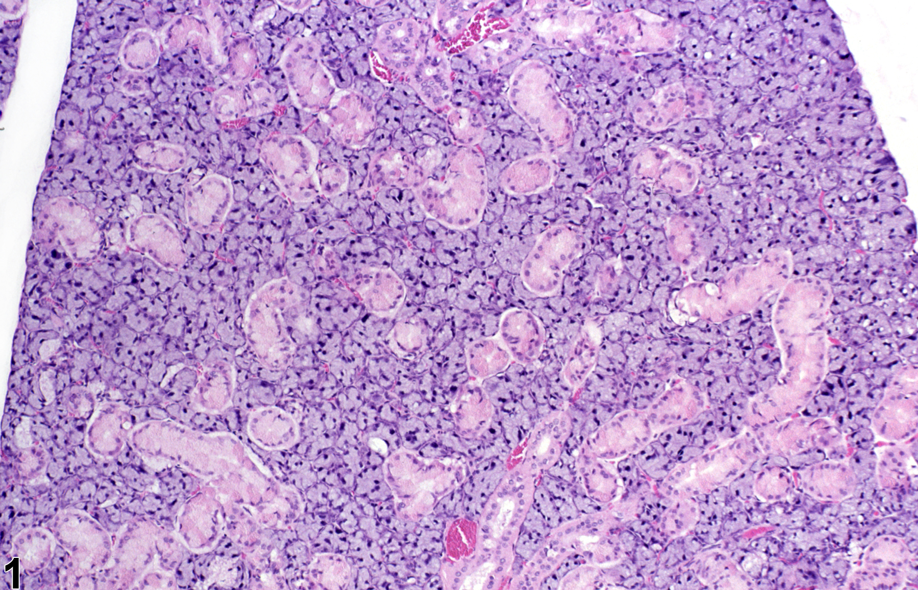
Normal submandibular salivary gland in a male F344/N rat from a subchronic study.
All Images

Normal submandibular salivary gland in a male F344/N rat from a subchronic study.

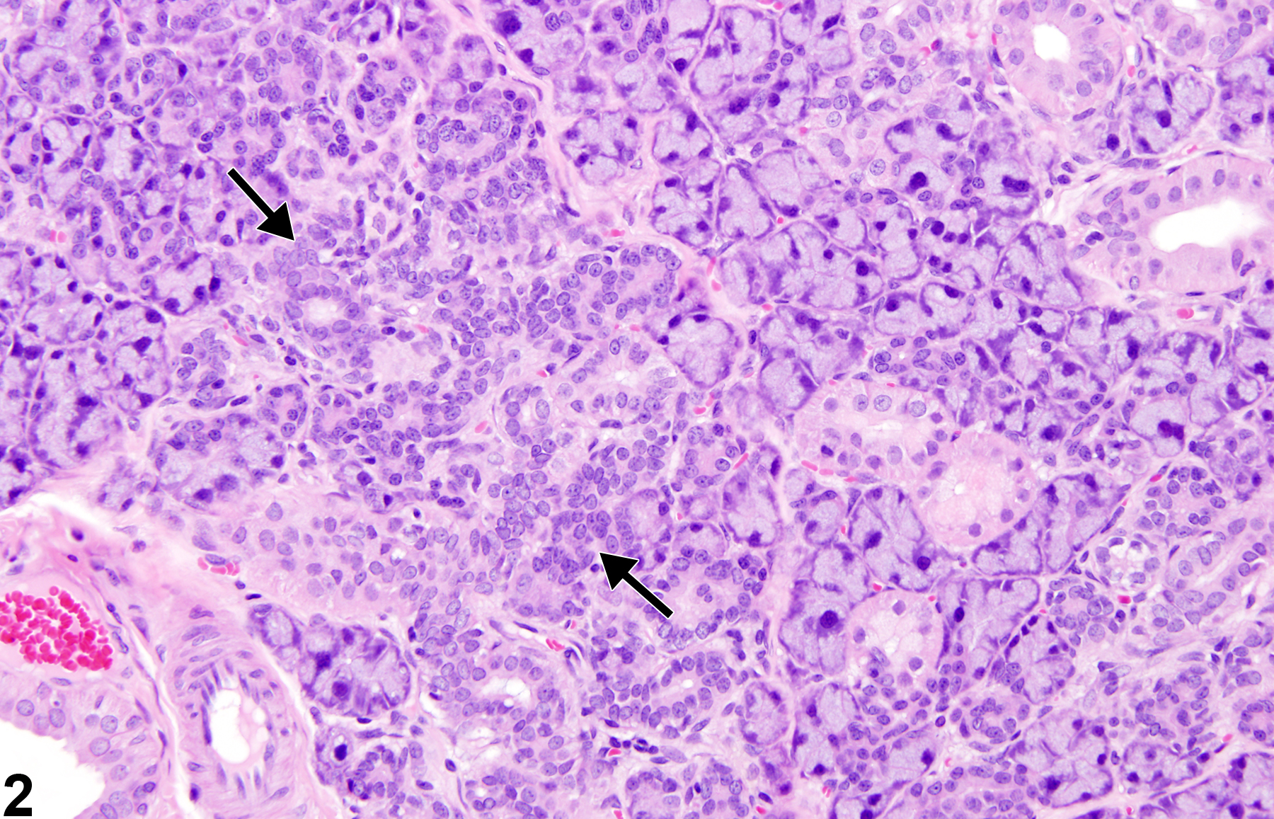
Salivary gland, Sublingual, Duct - Hyperplasia in a male F344/N rat from a subchronic study. There is an increase in the number of ductular epithelial cells within the gland (arrows).
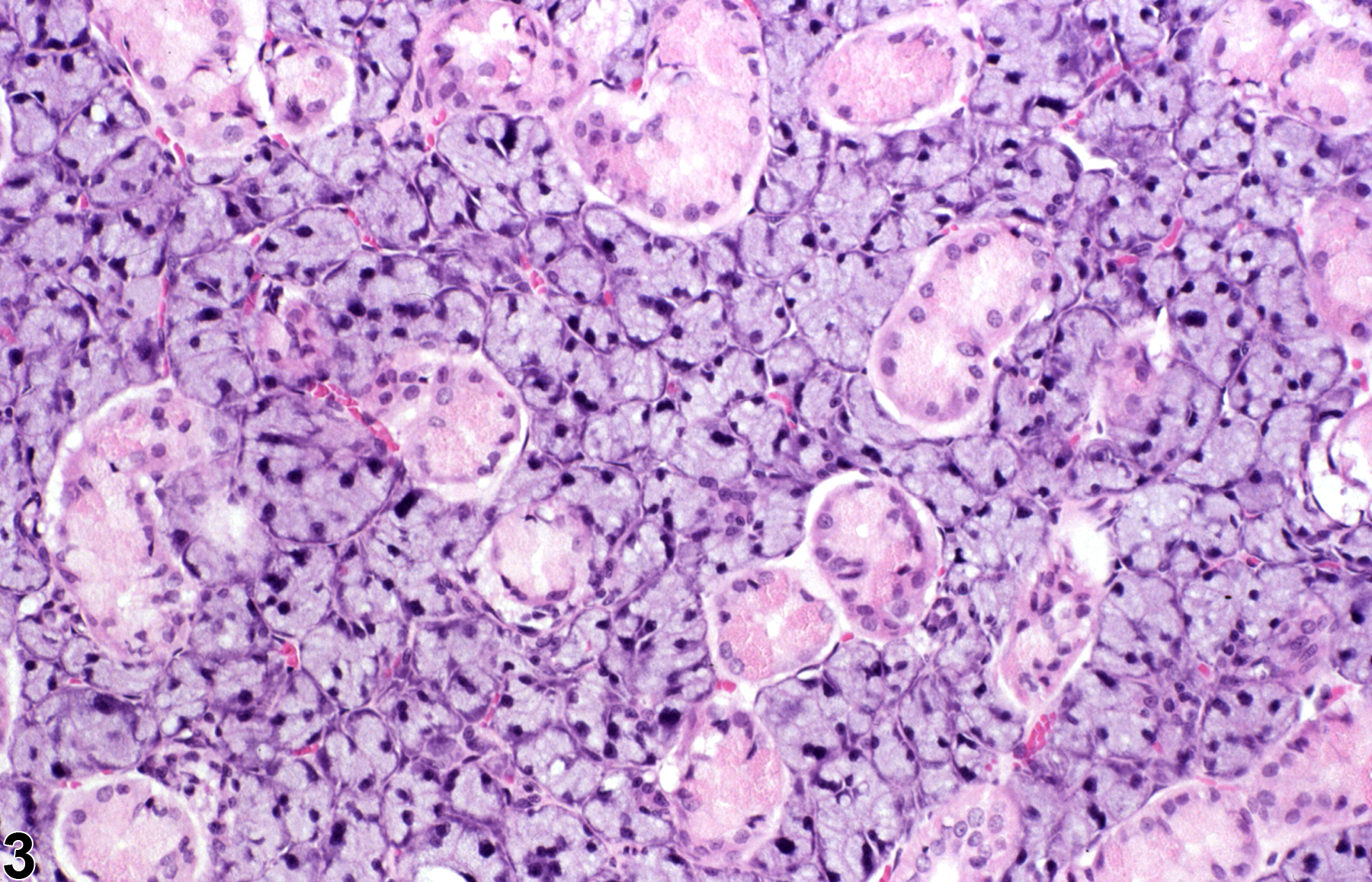
Normal salivary gland in a male F344/N rat from a subchronic study (higher magnification of Figure 1).

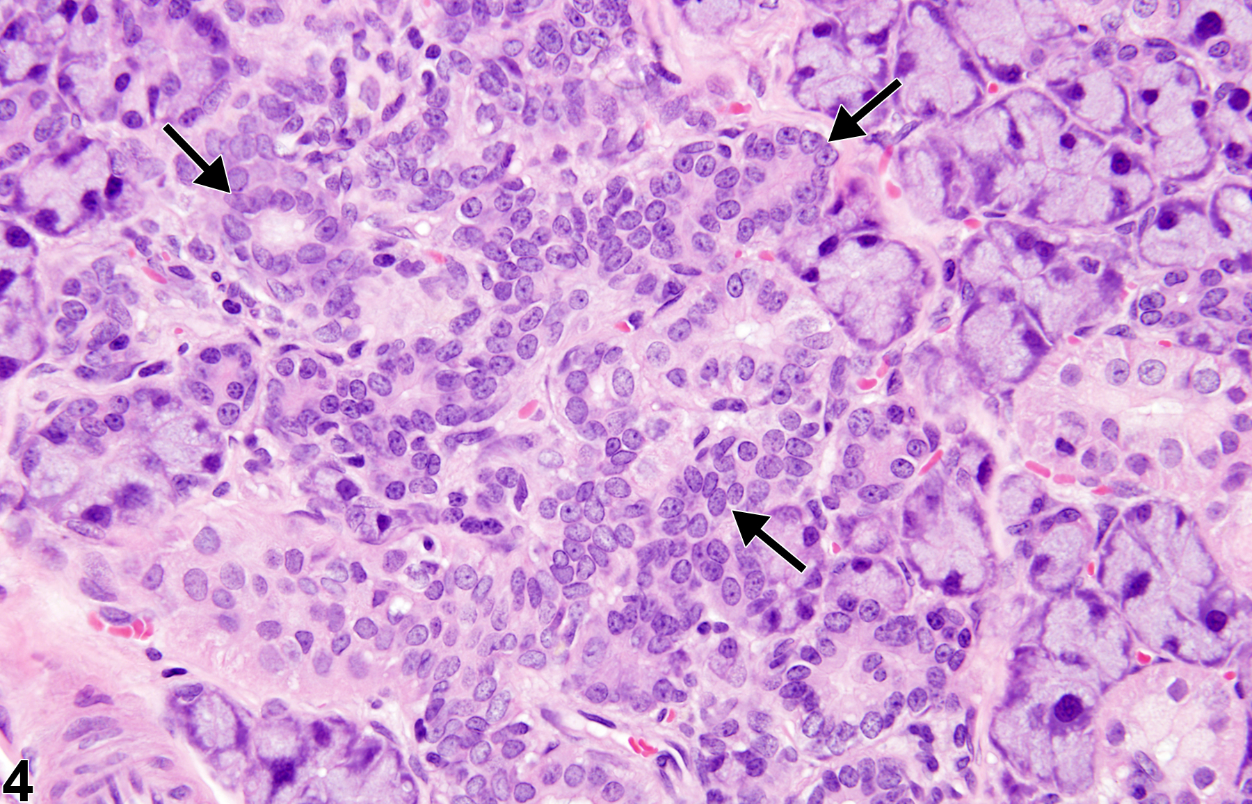
Salivary gland, Sublingual, Duct - Hyperplasia in a male F344/N rat from a subchronic study (higher magnification of Figure 2). There is an increase in the number of ductular epithelial cells within the gland (arrows).

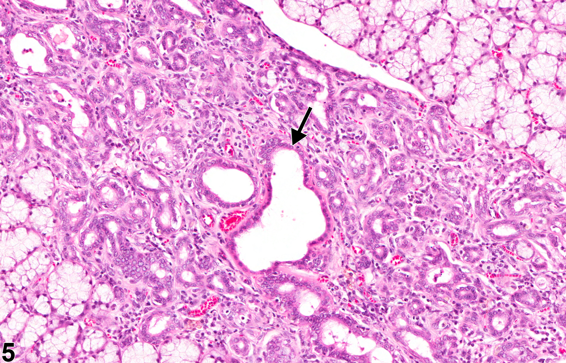
Salivary gland, Sublingual, Duct - Hyperplasia in a male F344/N rat from a chronic study. There is an increase in the number of ductal profiles within the gland. One duct is dilated and has an increased number of epithelial cells (arrow).

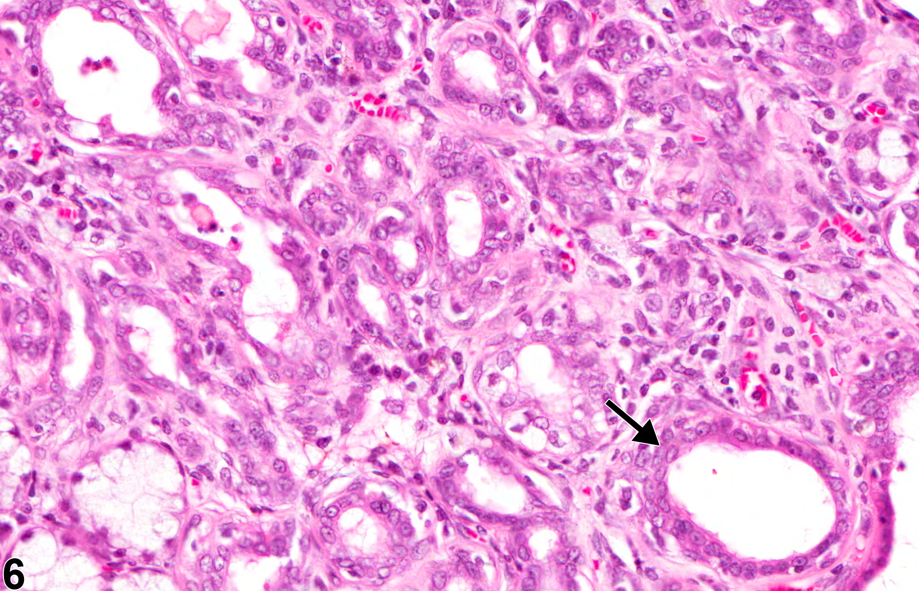
Salivary gland, Sublingual, Duct - Hyperplasia in a male F344/N rat from a chronic study (higher magnification of Figure 5). There is an increase in the number of ductal profiles within the gland. One duct is dilated and has an increased number of epithelial cells (arrow).