

Alimentary System
Salivary Gland - Infiltration, Cellular
Narrative
Botts S, Jokinen M, Gaillard ET, Elwell MR, Mann PC. 1999. Salivary, Harderian, and lacrimal glands. In: Pathology of the Mouse (Maronpot RR, ed). Cache River Press, St Louis, MO, 49-80.
Greaves P. 2007. Digestive system. In: Histopathology of Preclinical Toxicity Studies, 3rd ed. Academic Press, London, 334-456.
Neuenschwander SB, Elwell MR. 1990. Salivary glands. In: Pathology of the Fischer Rat (Boorman GA, Montgomery CA, MacKenzie WF, eds). Academic Press, San Diego, CA, 31-42.
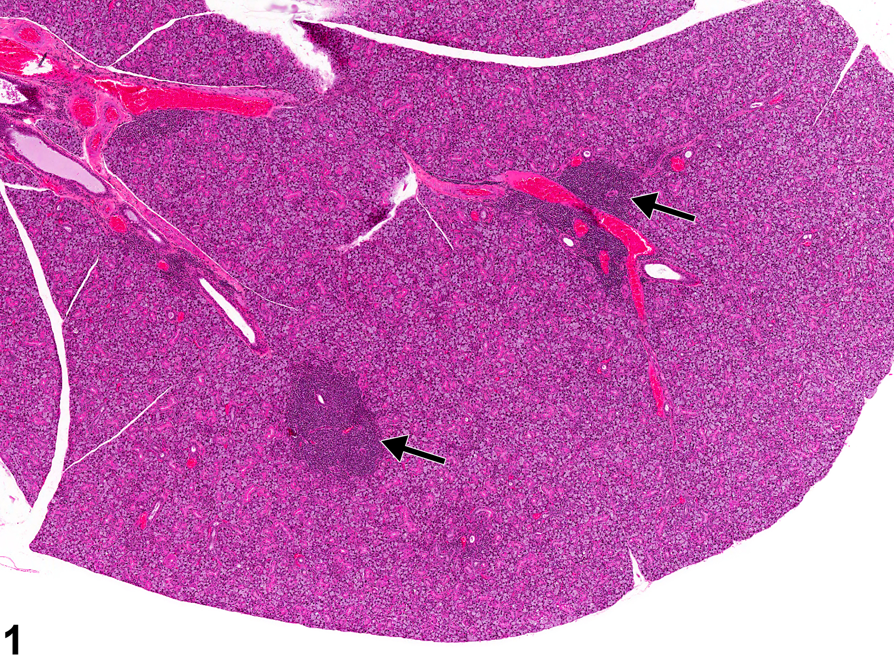
Salivary gland - Infiltration, Cellular, Lymphocyte in a female B6C3F1 mouse from a chronic study. The cellular infiltrates (arrows) are more deeply basophilic than the glandular tissue.
All Images

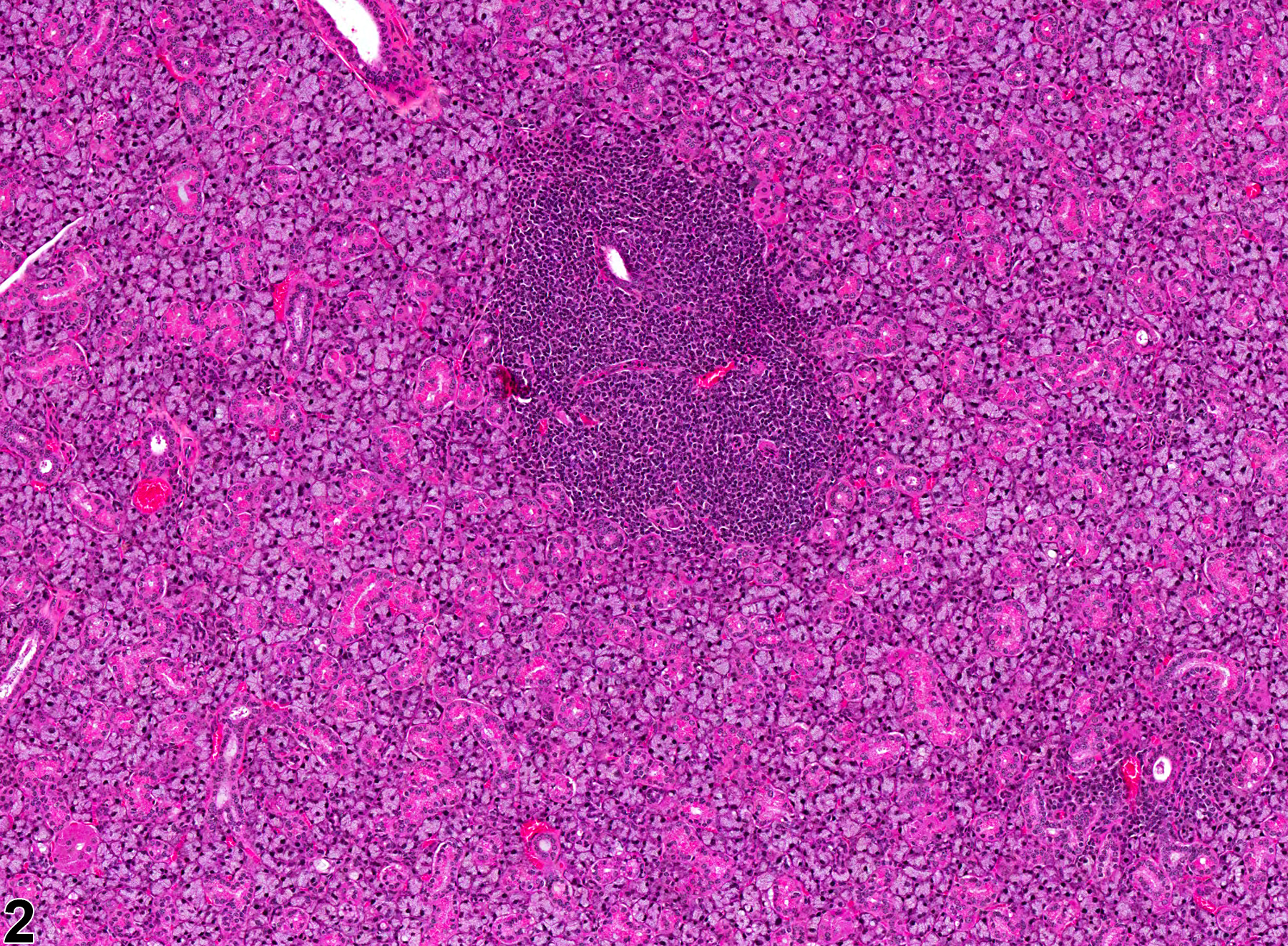
Salivary gland - Infiltration, Cellular, Lymphocyte in a female B6C3F1 mouse from a chronic study. The cellular infiltrates (arrows) are more deeply basophilic than the glandular tissue.

Salivary gland - Infiltration, Cellular, Lymphocyte in a female B6C3F1 mouse from a chronic study (higher magnification of Figure 1). The cellular infiltrates are composed mainly of lymphocytes.