U.S. flag

An official website of the United States government

Dot gov

The .gov means it's official.
Federal government websites often end in .gov or .mil. Before sharing sensitive information, make sure you're on a federal government site.

Https

The site is secure.
The https:// ensures that you are connecting to the official website and that any information you provide is encrypted and transmitted securely.

Skip to Main Navigation
Skip to Page Content
Skip to Atlas Navigation

Salivary Gland - Inflammation

Image of inflammation in the salivary gland from a male F344/N rat in a subchronic study
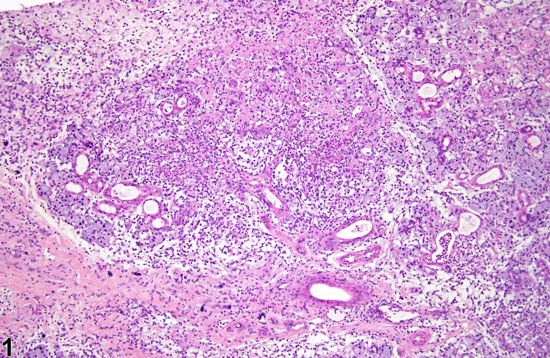
Salivary gland - Inflammation, Acute in a male F344/N rat from a subchronic study. There is widespread destruction of the salivary tissue with abundant inflammatory cells.
Figure 1 of 6
Image of inflammation in the salivary gland from a male F344/N rat in a subchronic study
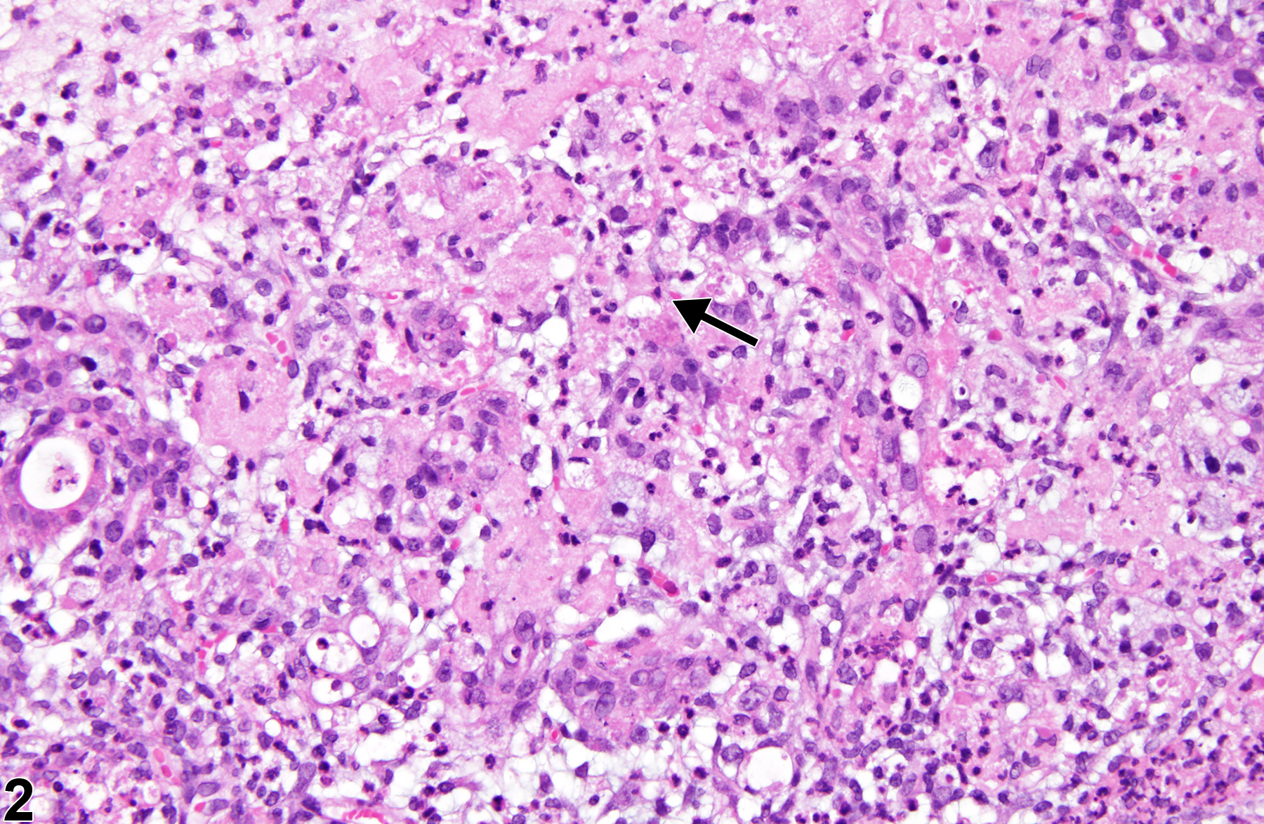
Salivary gland - Inflammation, Acute in a male F344/N rat from a subchronic study (higher magnification of Figure 1). The inflammatory cells are mainly neutrophils, and there is necrosis of the glandular tissue (arrow).
Figure 2 of 6
Image of inflammation in the salivary gland from a female B6C3F1 mouse in a chronic study
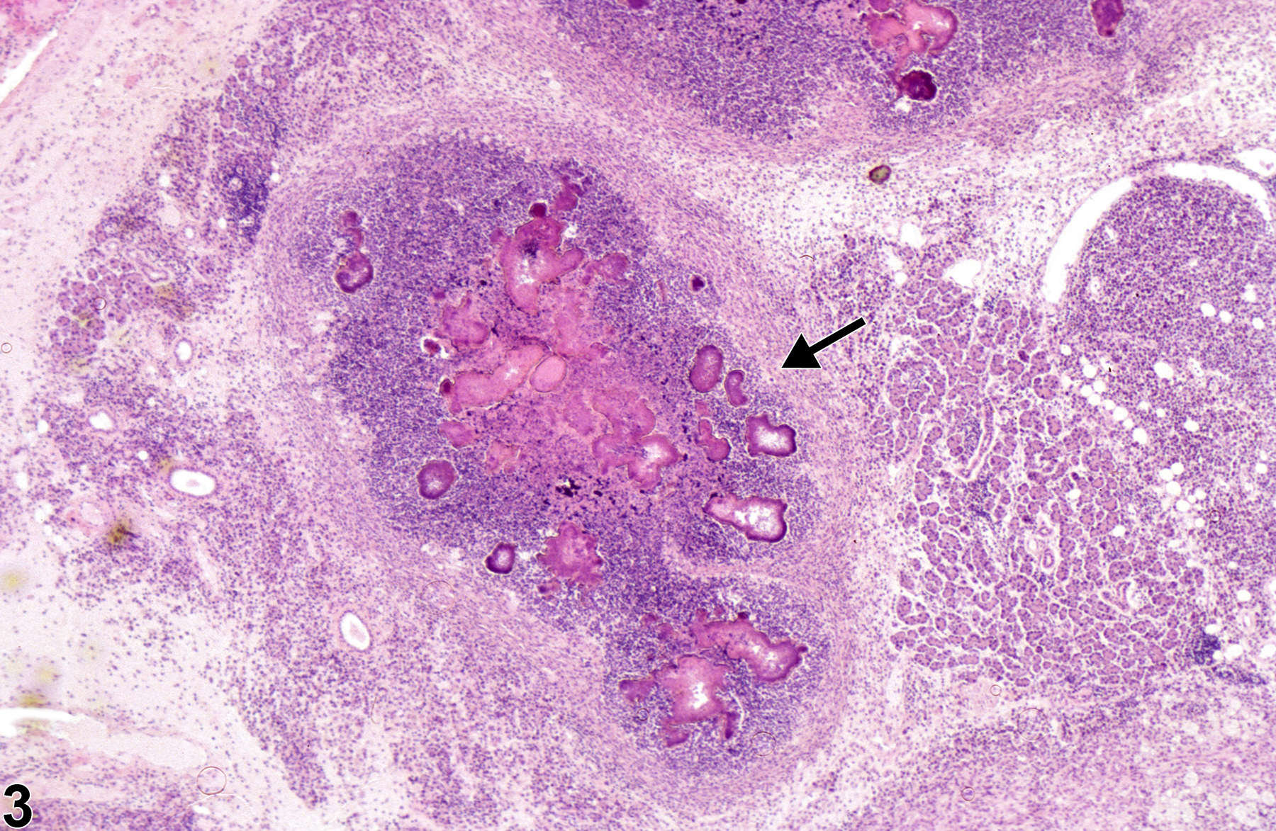
Salivary gland - Inflammation, Suppurative in a female B6C3F1 mouse from a chronic study. There are areas of inflammation with necrosis surrounded by fibrosis (arrow), characteristic of an abscess.
Figure 3 of 6
Image of inflammation in the salivary gland from a female B6C3F1 mouse in a chronic study
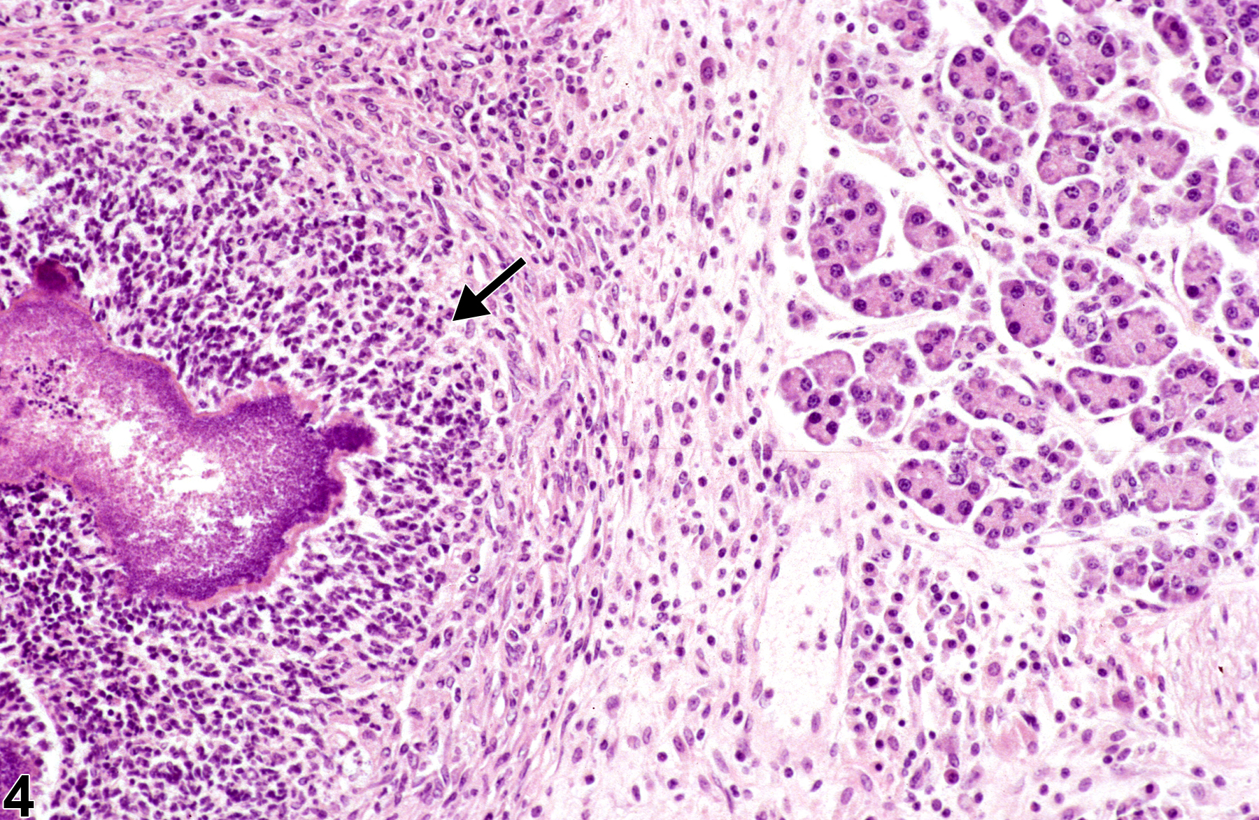
Salivary gland - Inflammation, Suppurative in a female B6C3F1 mouse from a chronic study (higher magnification of Figure 3). The inflammatory cells are mainly degenerate neutrophils surrounding large colonies of bacteria (arrow), which are surrounded by fibrosis.
Figure 4 of 6
Image of inflammation in the salivary gland from a male B6C3F1 mouse in a chronic study
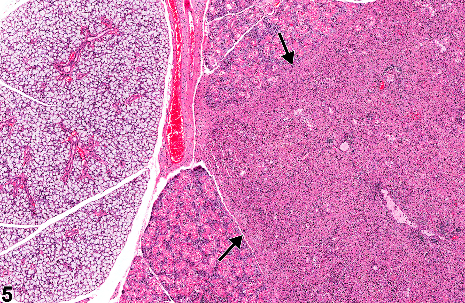
Salivary gland - Inflammation, Granulomatous in a male B6C3F1 mouse from a chronic study. An entire lobe is replaced by granulomatous inflammation (arrows).
Figure 5 of 6
Image of inflammation in the salivary gland from a male B6C3F1 mouse in a chronic study
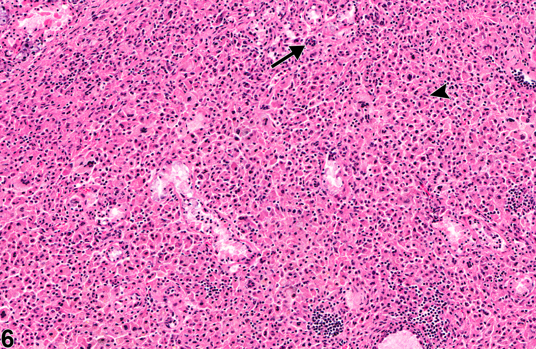
Salivary gland - Inflammation, Granulomatous in a male B6C3F1 mouse from a chronic study (higher magnification of Figure 5). The inflammation is characterized by the presence of multinucleated giant cells (arrow) and numerous epithelioid macrophages (arrowhead).
Figure 6 of 6
next arrow

comment:

In NTP studies, there are five standard categories of inflammation: acute ( Figure 1image opens in a pop-up window and Figure 2image opens in a pop-up window ), suppurative ( Figure 3image opens in a pop-up window and Figure 4image opens in a pop-up window ), chronic, chronic active, and granulomatous ( Figure 5image opens in a pop-up window and Figure 6image opens in a pop-up window ). In acute inflammation, the predominant infiltrating cell is the neutrophil, though fewer macrophages and lymphocytes may also be present. There may also be evidence of edema or hyperemia. The neutrophil is also the predominant infiltrating cell type in suppurative inflammation, but the neutrophils are aggregated, and many of them are degenerate (suppurative exudate). The exudate may contain cell debris, both from the resident cell populations and from infiltrating leukocytes; proteinaceous fluid containing fibrin; fewer macrophages; occasional lymphocytes or plasma cells; and, possibly, an infectious agent. Grossly, these lesions would be characterized by the presence of pus. The tissue surrounding the exudate may contain fibroblasts, fibrous connective tissue, and mixed inflammatory cells, depending on the chronicity of the lesion. Lymphocytes predominate in chronic inflammation. Lymphocytes also predominate in chronic active inflammation, but there are also a significant number of neutrophils. Both lesions may contain macrophages. Granulomatous inflammation is another form of chronic inflammation, but this diagnosis requires the presence of a significant number of aggregated, large, activated macrophages, epithelioid macrophages, or multinucleated giant cells.

Chronic inflammation can be seen in rats infected with sialodacryoadenitis corona virus, chronic infections with Klebsiella aerogenes, and immune-mediated sialoadenitis in mice. Sialodacryoadenitis virus (a corona virus) is the most important infectious agent affecting the salivary glands of rats (mice are not susceptible) because of its potential to compromise the interpretation of toxicologic studies. The virus, which mainly affects the submandibular and parotid salivary glands, causes gross enlargement of the glands, necrosis of both acinar and ductular epithelium, and marked inflammation. The sublingual salivary glands are not generally affected. In the acute phase, a neutrophilic infiltrate is associated with the necrosis, but later the infiltrates consist predominantly of mononuclear cells (chronic inflammation).

recommendation:

Whenever present, inflammation should be diagnosed and given a severity grade. A modifier indicating the duration or type of inflammation (i.e., acute, suppurative, chronic, chronic active, or granulomatous) should be included in the diagnosis. The severity grade depends on the extent of area of salivary gland affected and the density of the cellular infiltrate. Lesions consistent with an abscess are diagnosed as suppurative inflammation. Associated lesions, such as fibrosis, necrosis, edema, and hemorrhage, should not be diagnosed separately unless warranted by severity.

references:

Ackermann MR. 2007. Chronic inflammation and wound healing. In: Pathologic Basis of Veterinary Disease, 4th ed (McGavin MD, Zachary JF, eds). Mosby, St Louis, MO, 153-191.

Arseculeratne SN, Panabokke RG, Navaratnam C, Weliange LV. 1981. An epizootic of Klebsiella aerogenes infection in laboratory rats. Lab Anim 15:333-337.
Abstract: https://www.ncbi.nlm.nih.gov/pubmed/7043077

Botts S, Jokinen M, Gaillard ET, Elwell MR, Mann PC. 1999. Salivary, Harderian, and lacrimal glands. In: Pathology of the Mouse (Maronpot RR, ed). Cache River Press, St Louis, MO, 49-80.
Abstract: http://www.cacheriverpress.com/books/pathmouse.htm

Neuenschwander SB, Elwell MR. 1990. Salivary glands. In: Pathology of the Fischer Rat (Boorman GA, Montgomery CA, MacKenzie WF, eds). Academic Press, San Diego, CA, 31-42.
Abstract: https://www.ncbi.nlm.nih.gov/nlmcatalog/9002563