U.S. flag

An official website of the United States government

Dot gov

The .gov means it's official.
Federal government websites often end in .gov or .mil. Before sharing sensitive information, make sure you're on a federal government site.

Https

The site is secure.
The https:// ensures that you are connecting to the official website and that any information you provide is encrypted and transmitted securely.

Skip to Main Navigation
Skip to Page Content
Skip to Atlas Navigation

Salivary Gland, Parotid, Acinus - Hypertrophy

Image of normal comparison to hypertrophy in the parotid salivary gland acinus from a male F344/N rat in a subchronic study
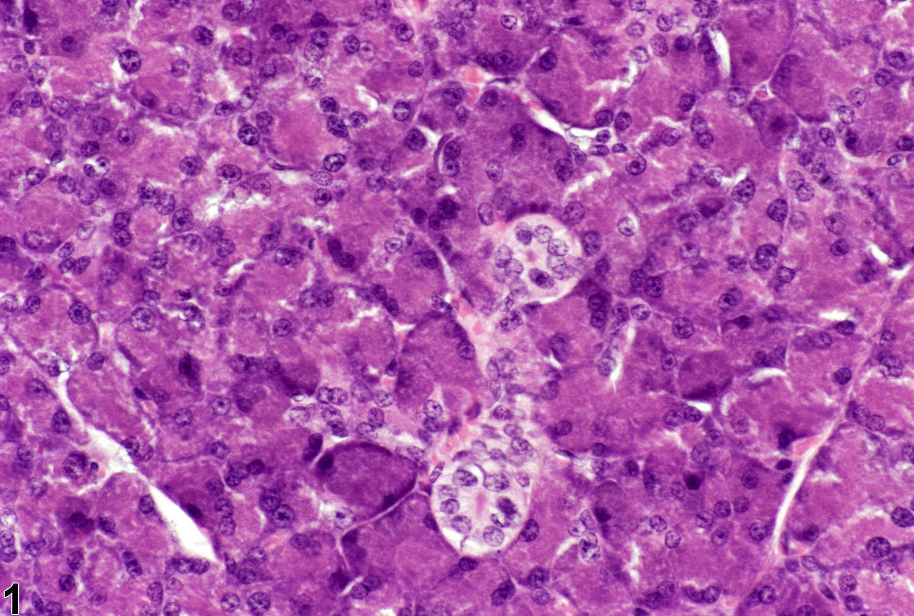
Normal parotid salivary gland from a male F344/N rat from a subchronic study.
Figure 1 of 2
Image of hypertrophy in the parotid salivary gland acinus from a male F344/N rat in a subchronic study
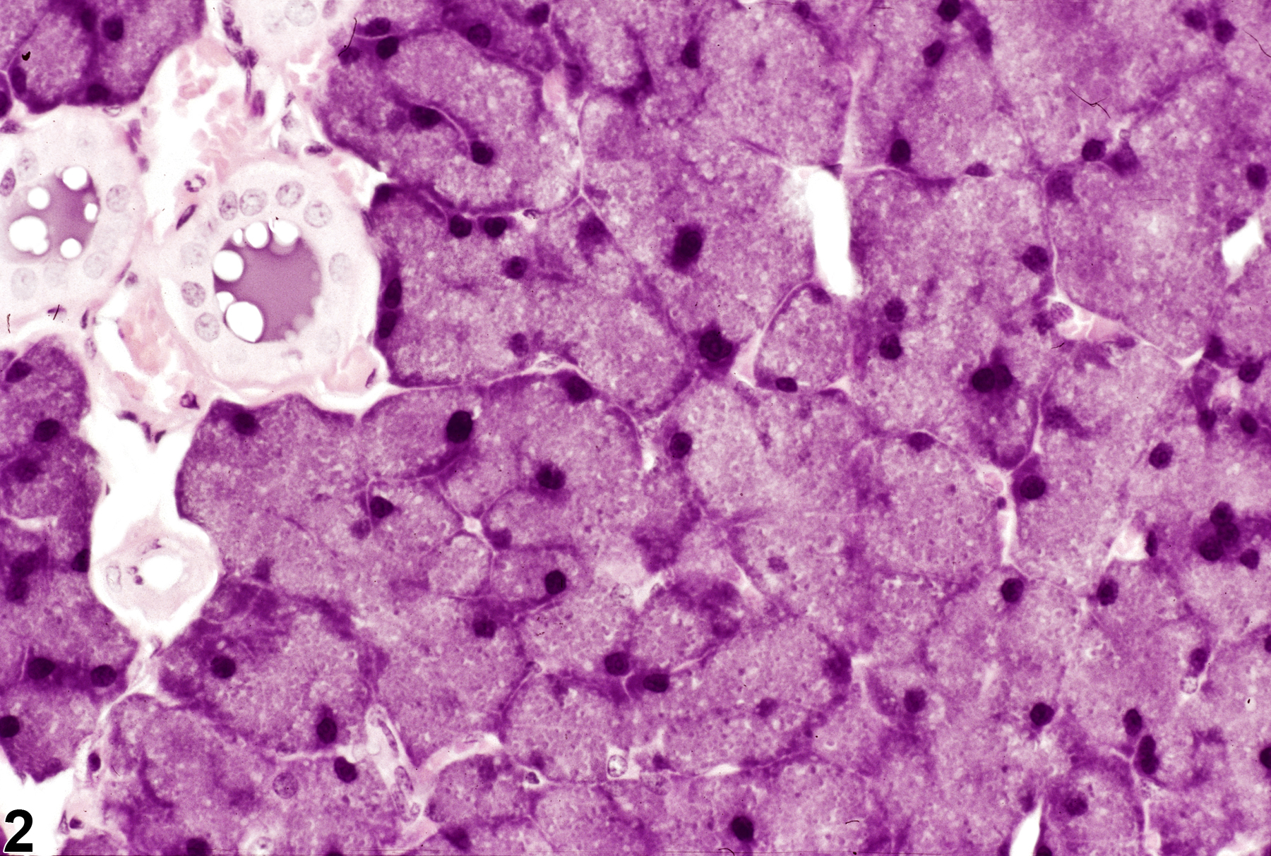
Salivary gland, Parotid, Acinus - Hypertrophy in a male F344/N rat from a subchronic study. The acinar cells are diffusely enlarged.
Figure 2 of 2
next arrow

comment:

Diffuse hypertrophy of parotid salivary gland acinar cells not associated with basophilic foci is rare in NTP studies. Likewise, diffuse acinar cell hypertrophy of the submandibular and sublingual salivary glands is rare in B6C3F1 mice and Fischer 344 rats. Acinar cell hypertrophy ( Figure 2image opens in a pop-up window ) is characterized by increased cell size primarily due to an increase in cytoplasm, although the nuclei are also usually larger in hypertrophied cells. The intense basophilia seen with hypertrophic basophilic foci is not present, and the cells are typically not as large. Diffuse hypertrophy of acinus cells often can be a subtle change requiring close comparison with control animals.

recommendation:

Salivary gland acinar hypertrophy should be diagnosed and graded based on the degree of increase in acinar cell size and number of cells affected. Multifocal hypertrophy would be assigned a higher severity grade than focal hypertrophy. If the hypertrophy is diffuse throughout the gland, then the modifier "diffuse" should be added to the diagnosis.

references:

Botts S, Jokinen M, Gaillard ET, Elwell MR, Mann PC. 1999. Salivary, Harderian, and lacrimal glands. In: Pathology of the Mouse (Maronpot RR, ed). Cache River Press, St Louis, MO, 49-80.
Abstract: http://www.cacheriverpress.com/books/pathmouse.htm

National Toxicology Program. 1992. NTP TOX-16. Toxicity Studies of Glyphosate (CAS No. 1071-83-6) Administered in Dosed Feed to F344/N Rats and B6C3F1 Mice. NTP, Research Triangle Park, NC.
Abstract: https://ntp.niehs.nih.gov/go/11968

Neuenschwander SB, Elwell MR. 1990. Salivary glands. In: Pathology of the Fischer Rat (Boorman GA, Montgomery CA, MacKenzie WF, eds). Academic Press, San Diego, CA, 31-42.
Abstract: https://www.ncbi.nlm.nih.gov/nlmcatalog/9002563