

Alimentary System
Salivary Gland, Submandibular - Secretory Depletion
Narrative
Comment:
A decrease in secretory product is seen occasionally and is often a subtle change requiring careful examination and comparison with controls to be detected. The cytoplasm of affected cells generally stains less intensely, and the cells are often smaller because of decreased amounts of cytoplasm. In the ductular cells, there is frequently a decrease in the number of eosinophilic, cytoplasmic granules. Secretory depletion differs from atrophy in that secretory depletion is a slight, diffuse change without an accompanying increase in ducts, interstitial tissue, or inflammatory cell infiltrate. Secretory depletion can occur in the acinar and ductular cells concurrently (Figure 2).
Recommendations:
Whenever present, secretory depletion should be diagnosed and graded based on the degree of decrease in secretory product in cells and number of cells involved. Secretory depletion in the convoluted ducts is based on degree of depletion of eosinophilic intracytoplasmic granules and number of convoluted (granular) ducts involved. If the secretory depletion is limited to the acinar cells, then no site modifier is necessary ("salivary gland, submandibular - secretory depletion" is diagnosed). If the change is limited to the ducts, then "salivary gland, submandibular, duct - secretory depletion" is diagnosed. If the change is present in both the acinar and ductal cells, then both diagnoses are made.
References:
Botts S, Jokinen M, Gaillard ET, Elwell MR, Mann PC. 1999. Salivary, Harderian, and lacrimal glands. In: Pathology of the Mouse (Maronpot RR, ed). Cache River Press, St Louis, MO, 49-80.
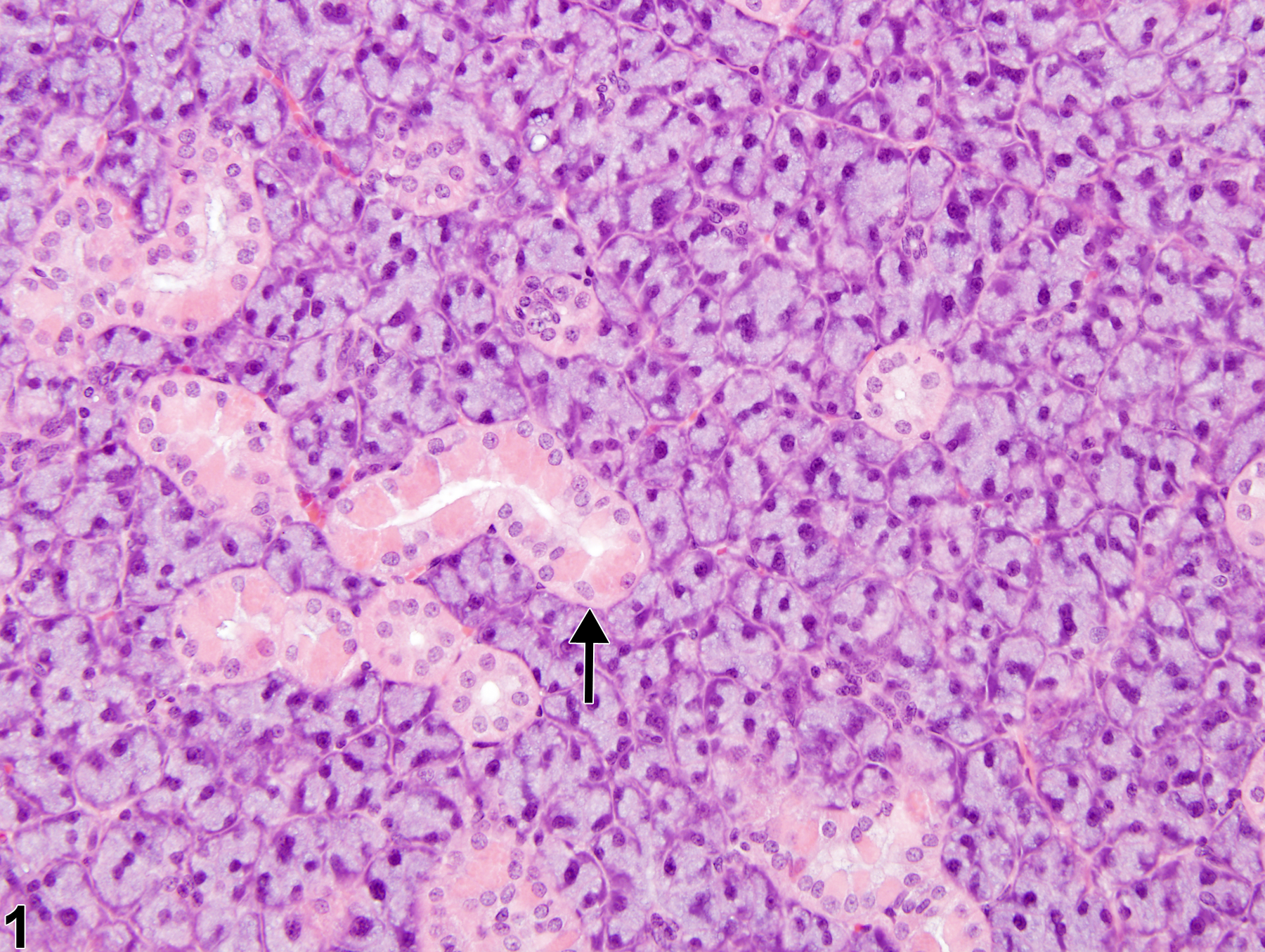
Normal submandibular salivary gland in a female F344/N rat from a subchronic study. The arrow identifies a convoluted duct.
All Images

Normal submandibular salivary gland in a female F344/N rat from a subchronic study. The arrow identifies a convoluted duct.

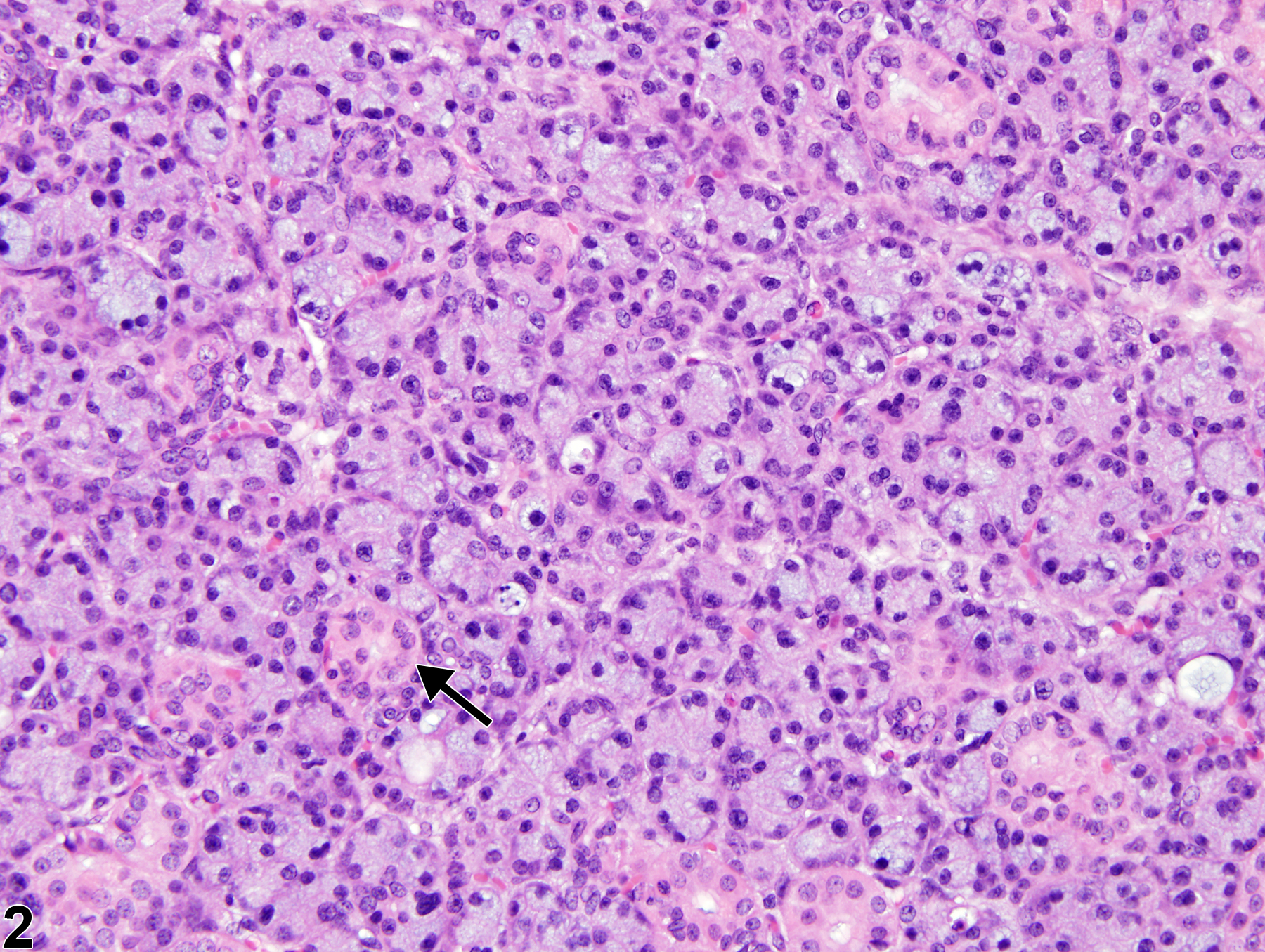
Salivary gland, Submandibular - Secretory depletion in a female F344/N rat from a subchronic study. The acinar and ductular (arrow) cells are smaller than those in Figure 1.