

Alimentary System
Tongue - Edema
Narrative
Comment:
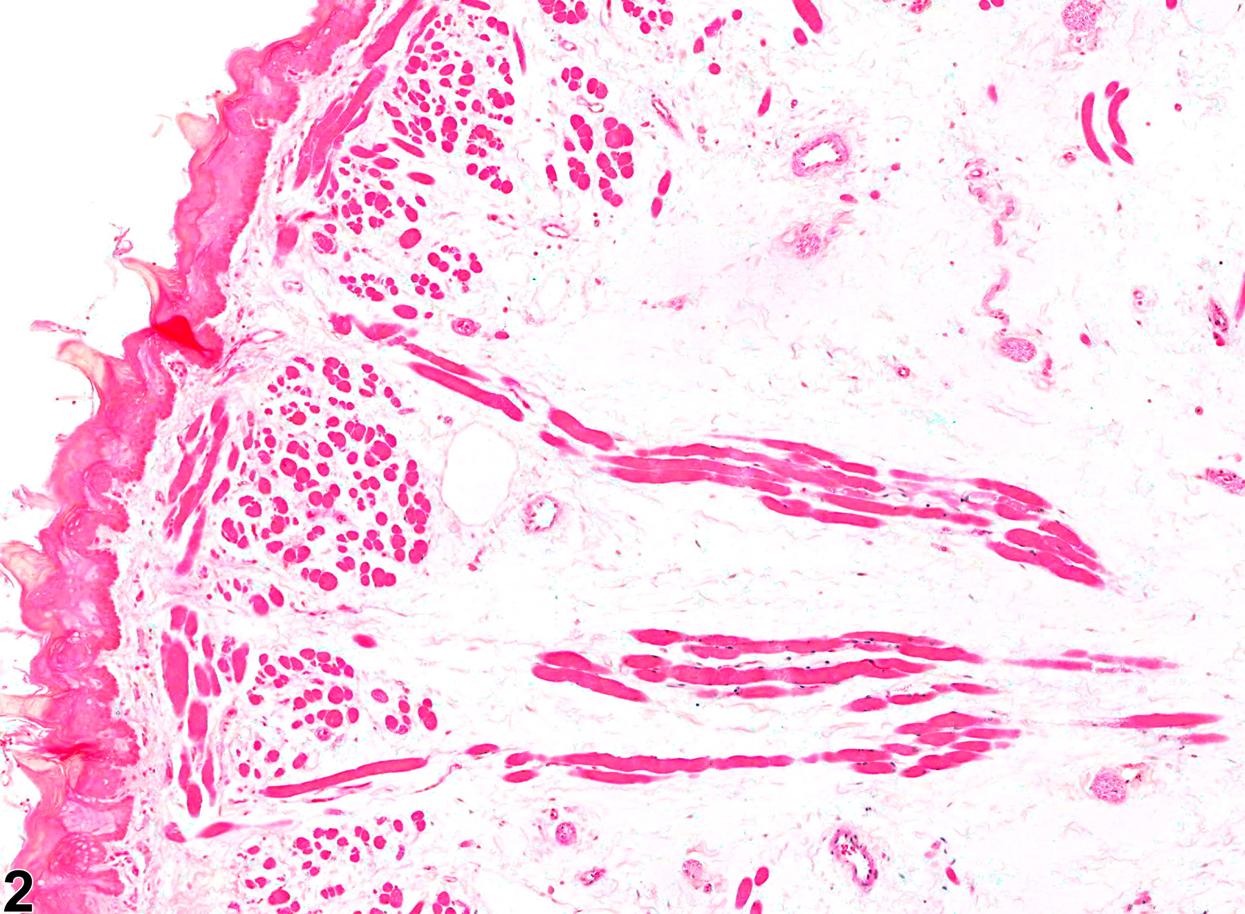
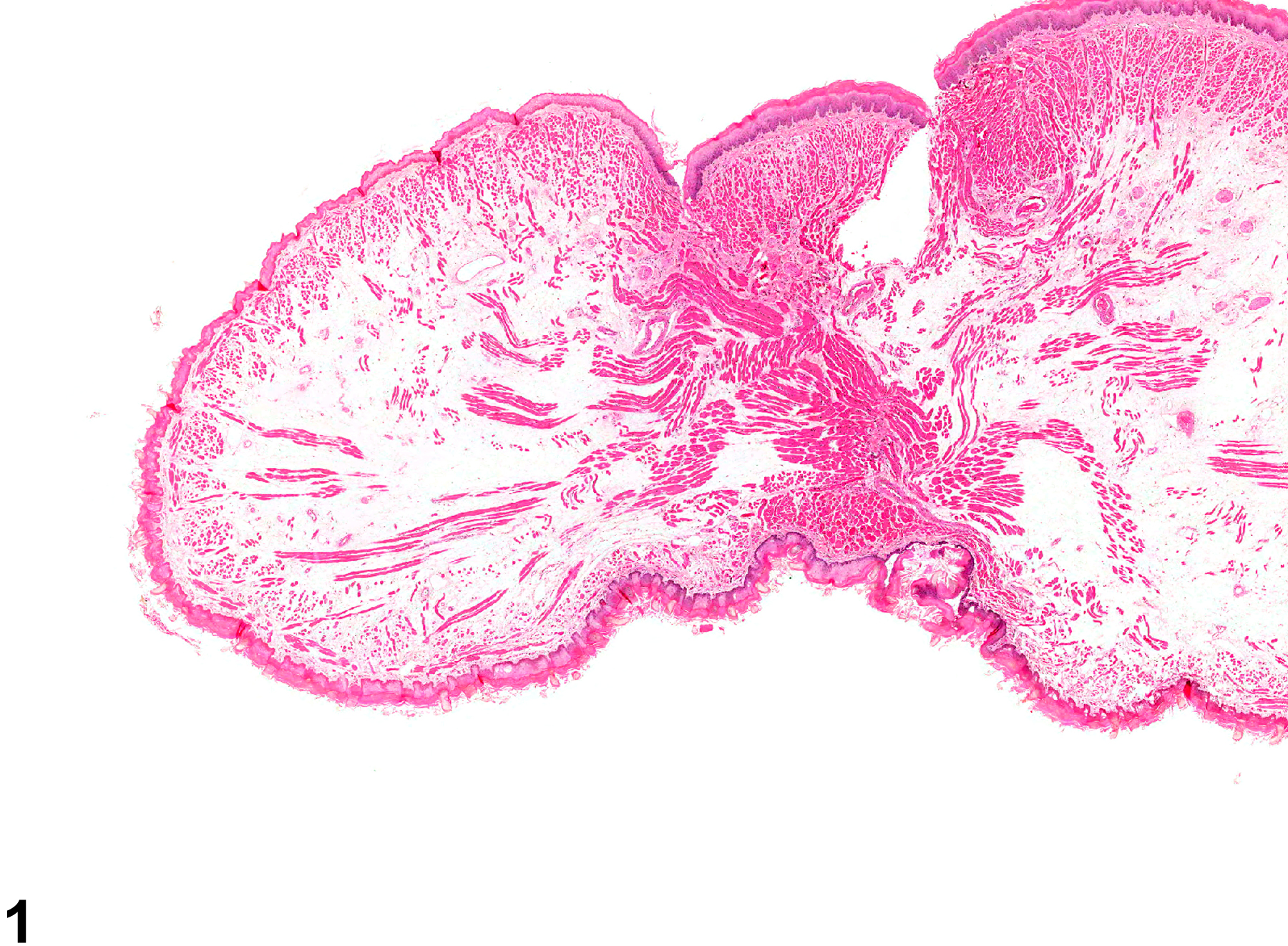
As in other organs, edema is the result of alteration in any of the factors that regulate the normal fluid distribution between the plasma, interstitium, and cells, such as increased vascular permeability, increased intravascular hydrostatic pressure, decreased intravascular osmotic pressure, and decreased lymphatic drainage. Therefore, any chemical that affects any of these factors can cause edema. Edema can also be a component of acute inflammation. Edema fluid appears as homogeneous, pale, eosinophilic material within the interstitium but is often lost in processing and appears as clear spaces (Figure 1 and Figure 2).
Recommendations:
Edema occurring in the absence of obvious inflammation should be diagnosed and graded based on the degree of separation of the tongue tissues by edema fluid and the total extent of tissue involvement. When it is associated with inflammation, it should be diagnosed separately unless it is very prominent or disproportionately severe, but it should be described in the pathology narrative as a component of the inflammation. Other associated lesions, such as necrosis, should be diagnosed and graded separately.
References:
Mosier DA. 2007. Vascular disorders and thrombosis. In: Pathologic Basis of Veterinary Disease, 4th ed (McGavin MD, Zachary JF, eds). Mosby, St Louis, MO, 63-99.
Tongue - Edema in a male F344/N rat from a chronic study. Edema in the tongue is evidenced by the clear spaces in the tissue.
All Images

Tongue - Edema in a male F344/N rat from a chronic study. Edema in the tongue is evidenced by the clear spaces in the tissue.

Tongue - Edema in a male F344/N rat from a chronic study (higher magnification of Figure 1). The edema is accompanied by necrosis of the associated epithelium.