

Alimentary System
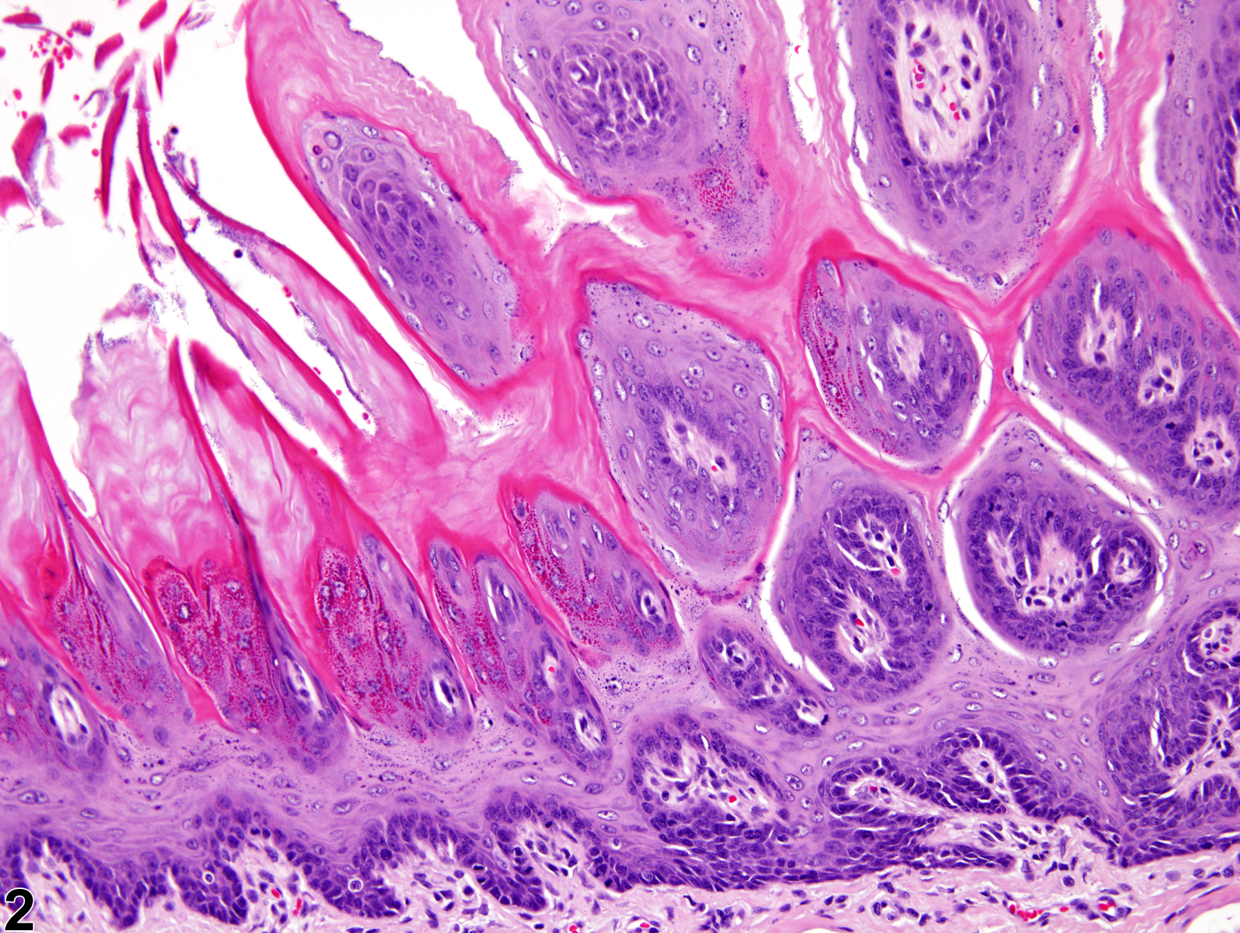
Tongue, Epithelium - Hyperplasia
Narrative
Brown HR, Hardisty JF. 1990. Oral cavity, esophagus and stomach. In: Pathology of the Fischer Rat (Boorman GA, Montgomery CA, MacKenzie WF, eds). Academic Press, San Diego, CA, 9-30.
Abstract: https://www.ncbi.nlm.nih.gov/nlmcatalog/9002563Leininger JR, Jokinen MP, Dangler CA, Whiteley LO. 1999. Oral cavity, esophagus, and stomach. In: Pathology of the Mouse (Maronpot RR, ed). Cache River Press, St Louis, MO, 29-48.
National Toxicology Program. 2010. NTP TR-544. Toxicology and Carcinogenesis Studies of Dibromoacetonitrile (CAS No. 3252-43-5) in F344/N Rats and B6C3F1 Mice (Drinking Water Studies). NTP, Research Triangle Park, NC.
Abstract: https://ntp.niehs.nih.gov/go/32617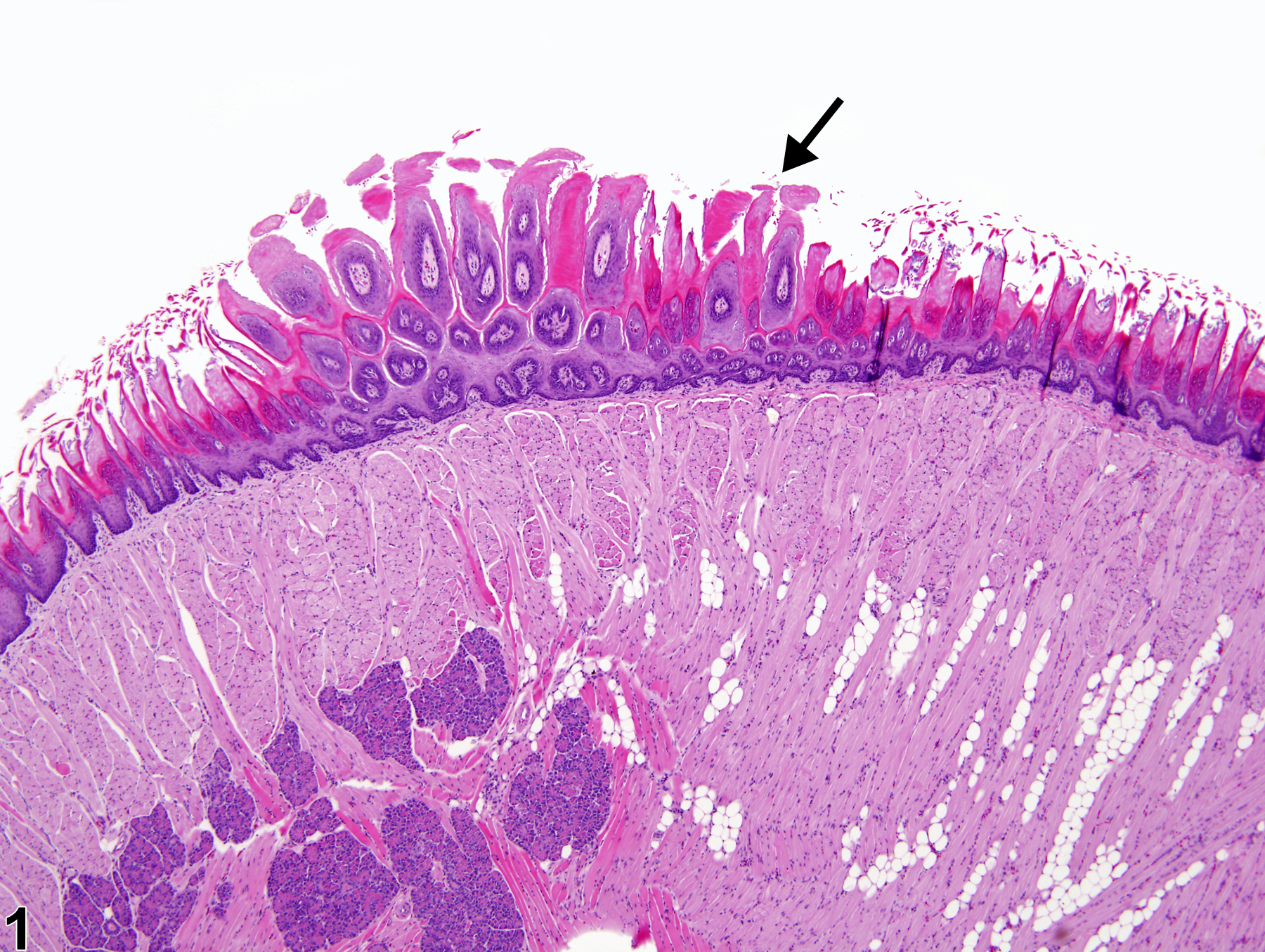
Tongue, Epithelium - Hyperplasia in a female F344/N rat from a chronic study. A focus of epithelial hyperplasia is present on the tongue (arrow).
All Images

Tongue, Epithelium - Hyperplasia in a female F344/N rat from a chronic study. A focus of epithelial hyperplasia is present on the tongue (arrow).

Tongue, Epithelium - Hyperplasia in a female F344/N rat from a chronic study (higher magnification of Figure 1). There are papillary epithelial projections in the focus of epithelial hyperplasia.