

Alimentary System
Tongue, Glands - Cyst
Narrative
Comment:
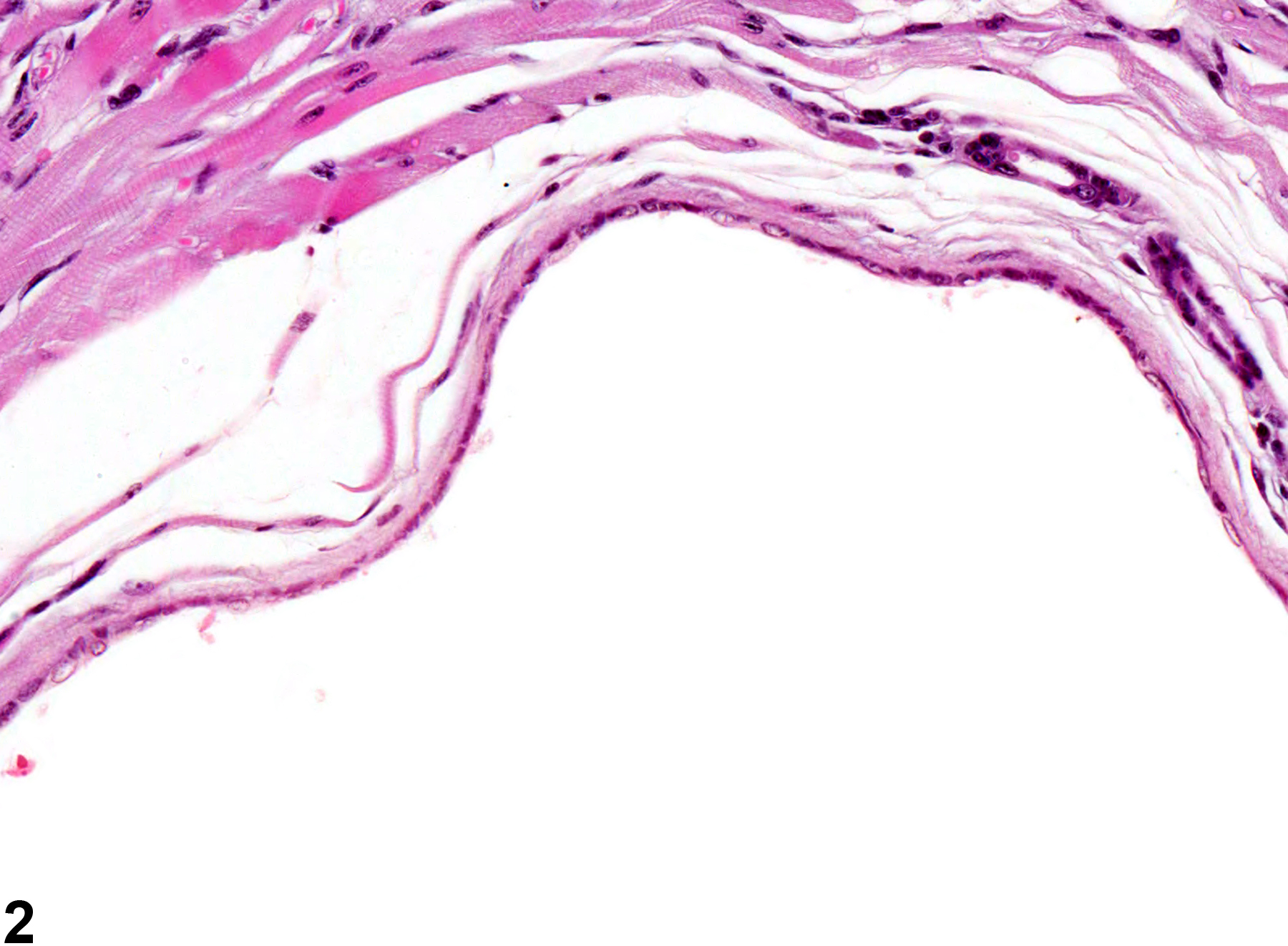
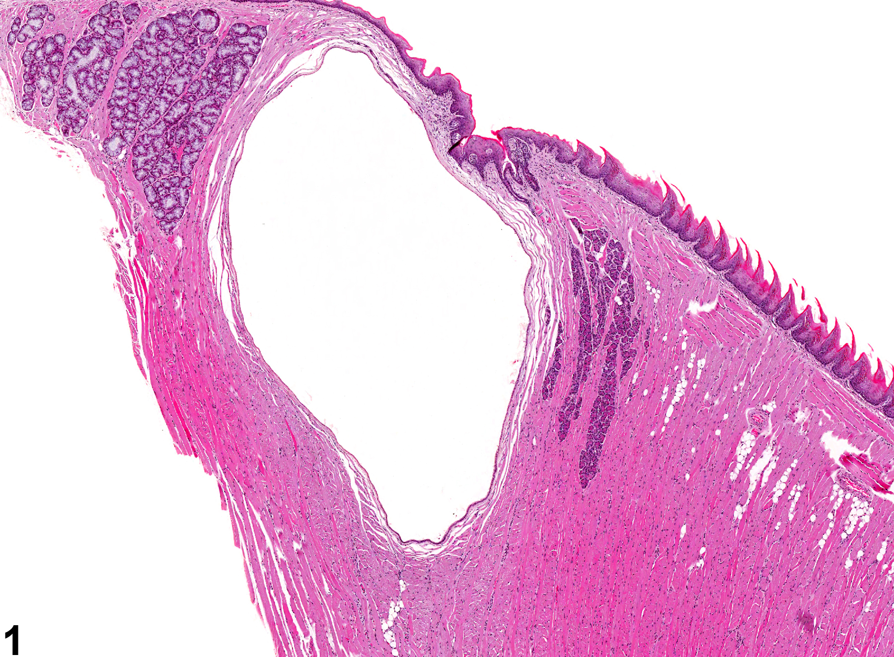
Cystic salivary ducts in the tongue (also known as ranulae) are lined by low cuboidal epithelium consistent with the salivary duct (as opposed to salivary mucoceles, which are cystic cavities filled with salivary secretions but not lined by an epithelium). Salivary gland duct cysts (Figure 1 and Figure 2) may be filled with pale eosinophilic fluid, but the fluid can be lost in processing, leaving an empty cyst. Cysts, dilations, or mucoceles may occur secondary to trauma, salivary calculi, or ductular foreign bodies and are considered incidental findings. Ruptured cysts may be accompanied by inflammation, often granulomatous. They are rare in F344/N rats and B6C3F1 mice.
Recommendations:
Whenever present, salivary duct cysts and dilations in the tongue should be diagnosed, but they are not generally graded. If the lesion is small, is diagnosed as dilation; larger lesions are diagnosed as cysts. If there is associated inflammation, the inflammation is not recorded separately unless warranted by severity.
References:
Botts S, Jokinen M, Gaillard ET, Elwell MR, Mann PC. 1999. Salivary, Harderian, and lacrimal glands. In: Pathology of the Mouse (Maronpot RR, ed). Cache River Press, St Louis, MO, 49-80.
Neuenschwander SB, Elwell MR. 1990. Salivary glands. In: Pathology of the Fischer Rat (Boorman GA, Montgomery CA, MacKenzie WF, eds). Academic Press, San Diego, CA, 31-42.
Tongue, Gland - Cyst in a female B6C3F1 mouse from a chronic study. There is a large cyst amid the salivary tissue in the tongue.
All Images

Tongue, Gland - Cyst in a female B6C3F1 mouse from a chronic study. There is a large cyst amid the salivary tissue in the tongue.

Tongue, Gland - Cyst in a female B6C3F1 mouse from a chronic study. The cyst is lined by flattened, nonkeratinized epithelium.