

Alimentary System
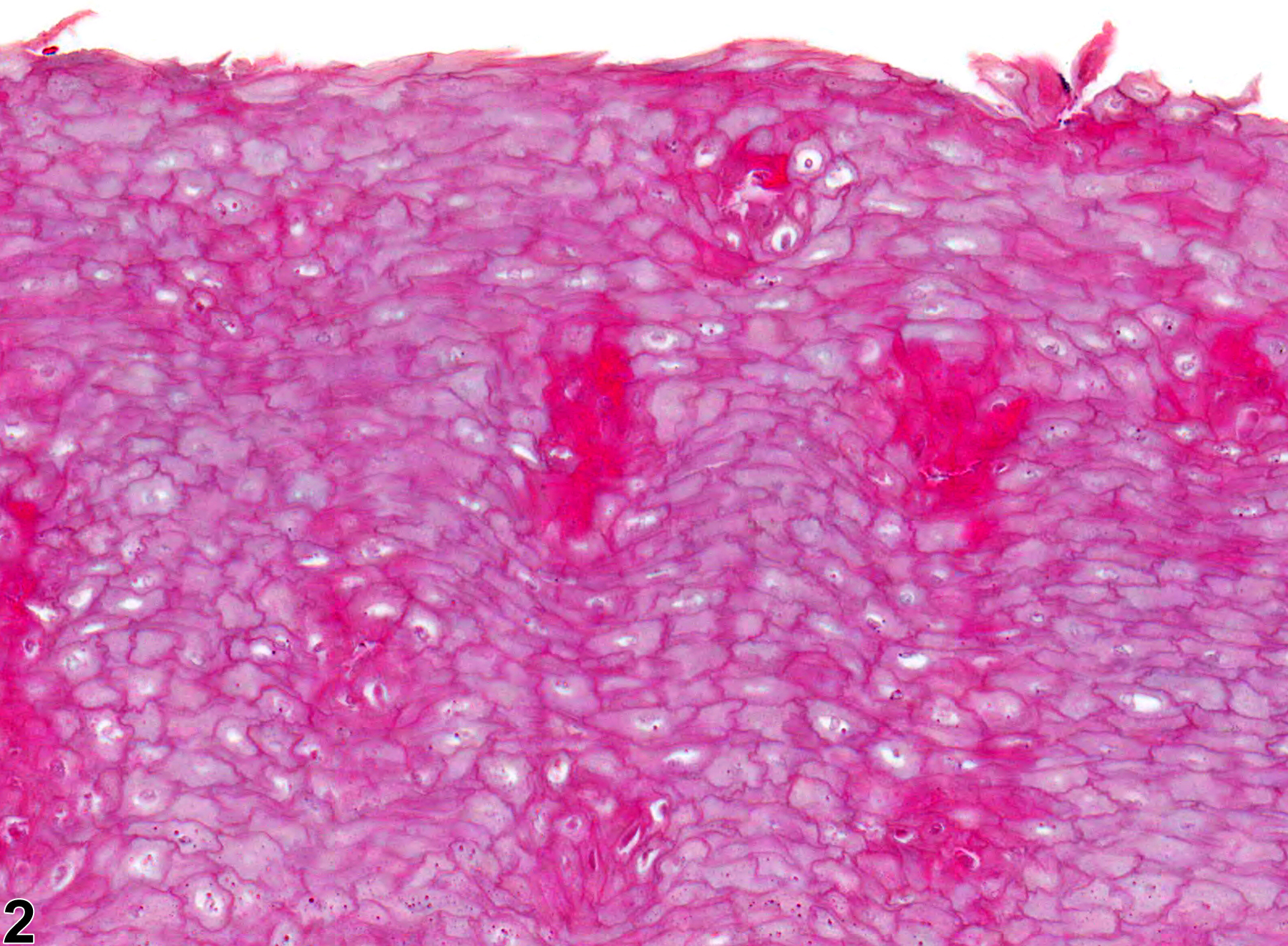
Tongue - Hyperkeratosis
Narrative
Hargis AM, Ginn PE. 2007. The integument. In: Pathologic Basis of Veterinary Disease, 4th ed (McGavin MD, Zachary JF, eds). Mosby, St Louis, MO, 1107-1261.
Kaplan I, Hochstadt T, Dayan D. 2002. PCNA in palate and tongue mucosal dysplastic lesions induced by topically applied 4NQO in desalivated rat. Med Pathol 7:336-343.
Abstract: https://www.ncbi.nlm.nih.gov/pubmed/12415217Leininger JR, Jokinen MP, Dangler CA, Whiteley LO. 1999. Oral cavity, esophagus, and stomach. In: Pathology of the Mouse (Maronpot RR, ed). Cache River Press, St Louis, MO, 29-48.
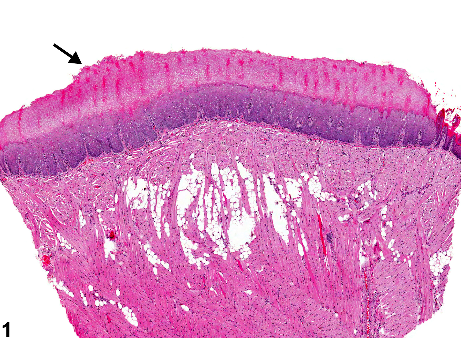
Tongue - Hyperkeratosis in a female F344/N rat from a chronic study. The keratin layer on the surface of the tongue is thickened (arrow).
All Images

Tongue - Hyperkeratosis in a female F344/N rat from a chronic study. The keratin layer on the surface of the tongue is thickened (arrow).

Tongue - Hyperkeratosis in a female F344/N rat from a chronic study (higher magnification of Figure 1). The hyperkeratosis is orthokeratotic, lacking nuclei.