U.S. flag

An official website of the United States government

Dot gov

The .gov means it's official.
Federal government websites often end in .gov or .mil. Before sharing sensitive information, make sure you're on a federal government site.

Https

The site is secure.
The https:// ensures that you are connecting to the official website and that any information you provide is encrypted and transmitted securely.

Skip to Main Navigation
Skip to Page Content
Skip to Atlas Navigation

Tongue - Ulcer

Image of ulcer in the tongue from a female F344/N rat in a chronic study
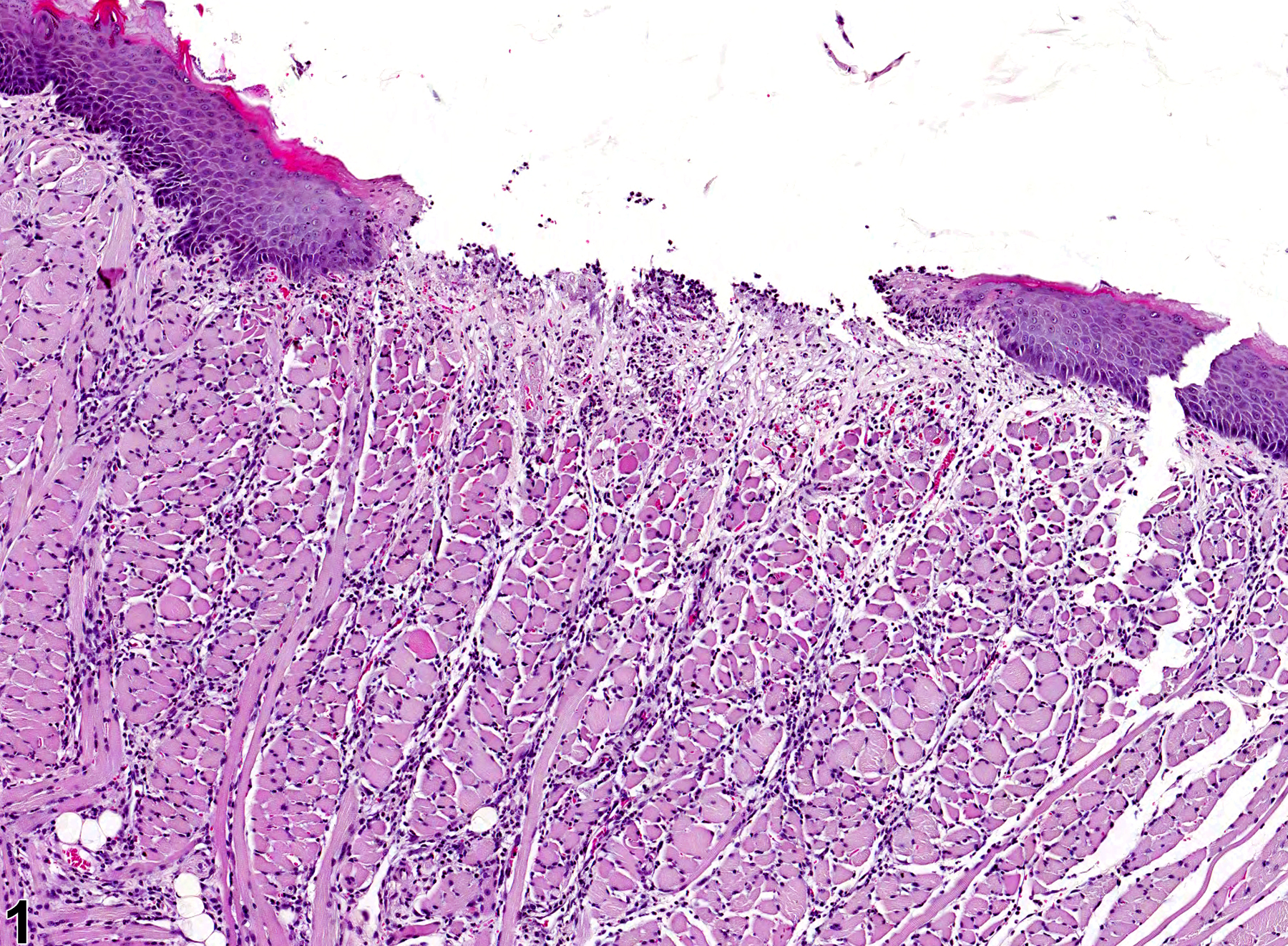
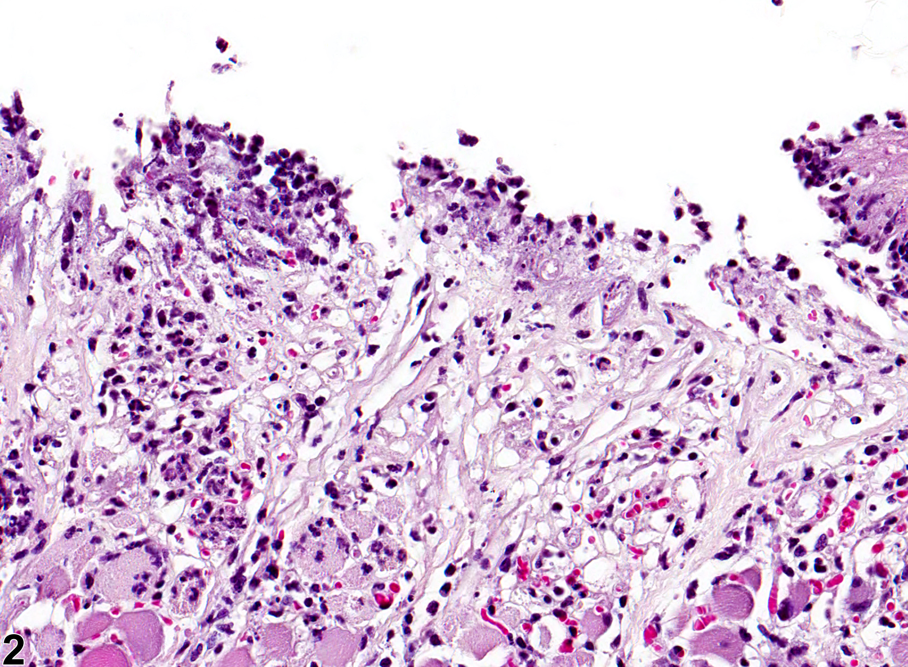
Tongue - Ulcer in a female F344/N rat from a chronic study. A portion of the keratinized squamous epithelium of the tongue has been lost, exposing the underlying lamina propria.
Figure 1 of 2
Image of ulcer in the tongue from a female F344/N rat in a chronic study
Tongue - Ulcer in a female F344/N rat from a chronic study (higher magnification of Figure 1). Inflammatory cells are present on the surface of the ulcer and within the underlying lamina propria.
Figure 2 of 2
next arrow

comment:

Ulcers and erosions can occur anywhere in the gastrointestinal tract, including the tongue. An erosion involves the loss of superficial epithelium of the mucosa, whereas an ulcer ( Figure 1image opens in a pop-up window and Figure 2image opens in a pop-up window ) involves loss of the full thickness of epithelial cells and extends to the lamina propria. Ulceration and inflammation can be the result of spontaneous disease or local or systemic toxic injury. Ulcers on the tongue can be caused by malocclusion with incisor overgrowth or can be secondary to uremia. They are typically accompanied by inflammation and may also be accompanied by myocyte degeneration.

recommendation:

Ulceration and erosion of the tongue should be diagnosed and graded. Edema, inflammation, and hyperplasia of the adjacent epithelium should not generally be diagnosed separately unless they are prominent components of the lesion. Necrosis of the epithelium is diagnosed instead of erosion or ulceration if the necrotic epithelium is still present and attached to the underlying lamina propria.

references:

Bertram TA, Markovits JE, Juliana MM. 1996. Non-proliferative lesions of the alimentary canal in rats GI-1. In: Guides for Toxicologic Pathology. STP/ARP/AFIP, Washington, DC, 1-16.
Full Text: https://www.toxpath.org/docs/SSNDC/GINonproliferativeRat.pdf

Comereski CR, Peden WM, Davidson TJ, Warner GL, Hirth RS, Frantz JD. 1994. BR96-doxorubicin conjugate (BMS-182248) versus doxorubicin: A comparative toxicity assessment in rats. Toxicol Pathol 22:473-488.
Abstract: https://www.ncbi.nlm.nih.gov/pubmed/7899776

Leininger JR, Jokinen MP, Dangler CA, Whiteley LO. 1999. Oral cavity, esophagus, and stomach. In: Pathology of the Mouse (Maronpot RR, ed). Cache River Press, St Louis, MO, 29-48.
Abstract: http://www.cacheriverpress.com/books/pathmouse.htm

National Toxicology Program. 2008. NTP TR-546. Toxicology and Carcinogenesis Studies of Sodium Dichromate Dihydrate (CAS No. 7789-12-0) in F344 rats and B6C3F1 Mice (Drinking Water Studies). NTP, Research Triangle Park, NC.
Abstract: https://ntp.niehs.nih.gov/go/29323