U.S. flag

An official website of the United States government

Dot gov

The .gov means it's official.
Federal government websites often end in .gov or .mil. Before sharing sensitive information, make sure you're on a federal government site.

Https

The site is secure.
The https:// ensures that you are connecting to the official website and that any information you provide is encrypted and transmitted securely.

Skip to Main Navigation
Skip to Page Content
Skip to Atlas Navigation

Tooth, Ameloblast - Atrophy

Image of normal ameloblast comparison to atrophy in the tooth from a male B6C3F1 mouse in a chronic study
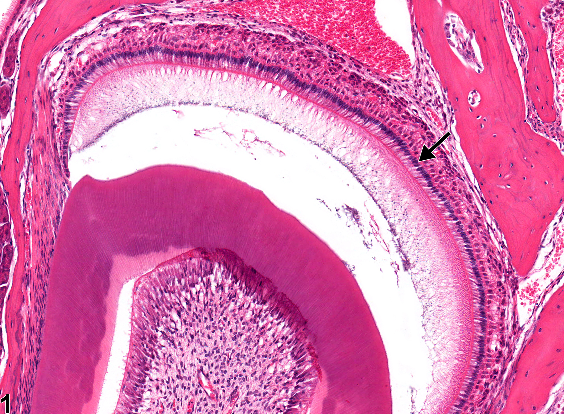
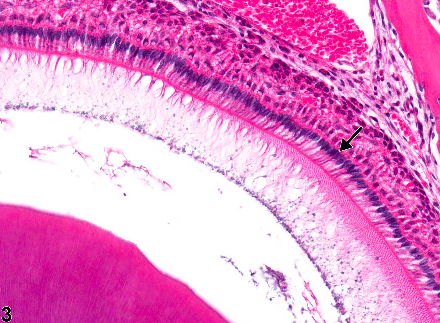
Normal ameloblasts (arrow) in a male B6C3F1 mouse from a chronic study.
Figure 1 of 4
Image of atrophy in the tooth ameloblast from a male F344/N rat in a chronic study
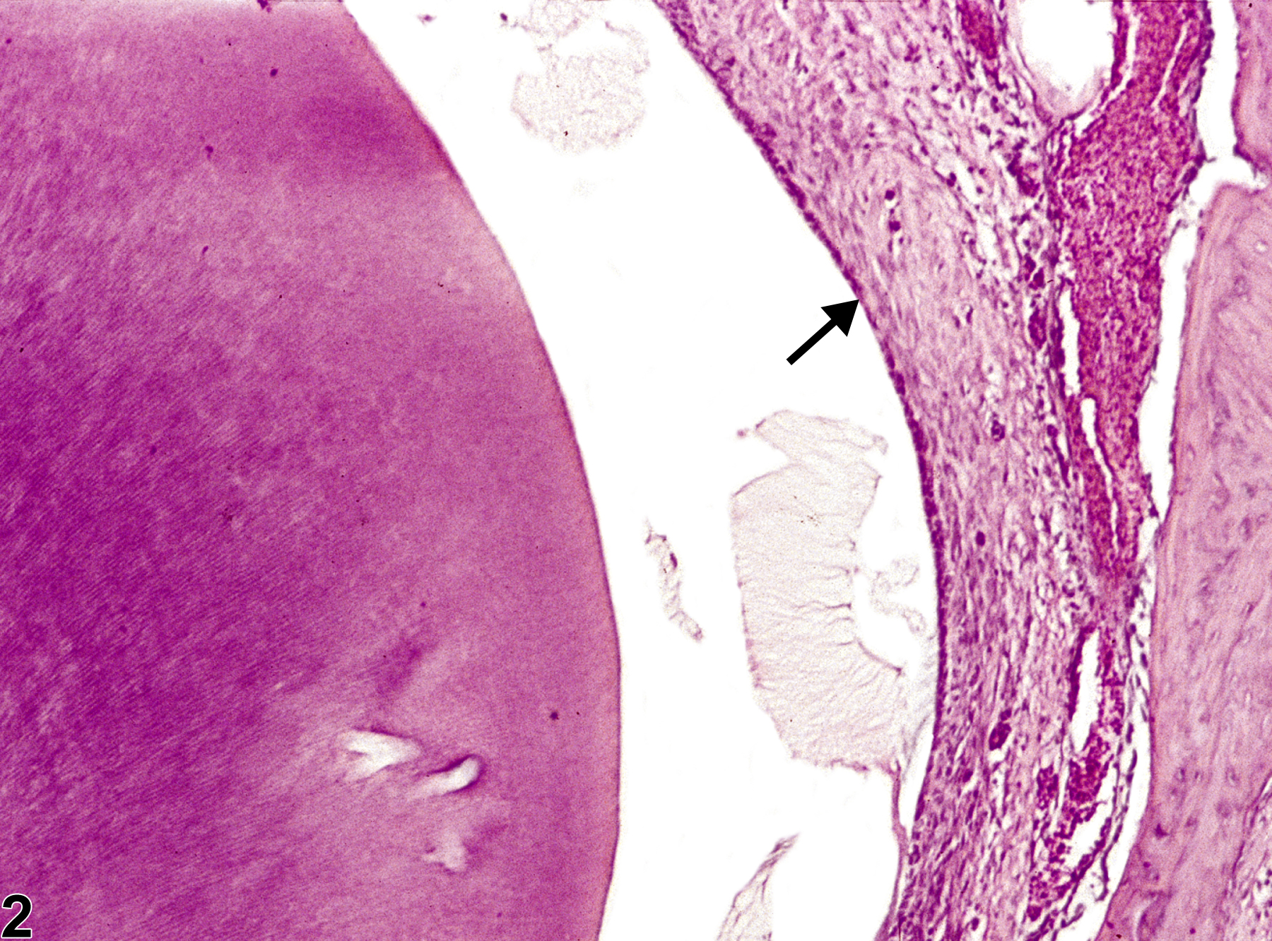
Tooth, Ameloblast - Atrophy in a male F344/N rat from a chronic study. The ameloblast layer is thin (compare with Figure 1), due to the decreased size of the ameloblasts (arrow).
Figure 2 of 4
Image of normal ameloblast comparison to atrophy in the tooth from a male B6C3F1 mouse in a chronic study
Normal ameloblasts (arrow) in a male B6C3F1 mouse from a chronic study (higher magnification of Figure 1).
Figure 3 of 4
Image of atrophy in the tooth ameloblast from a male F344/N rat in a chronic study
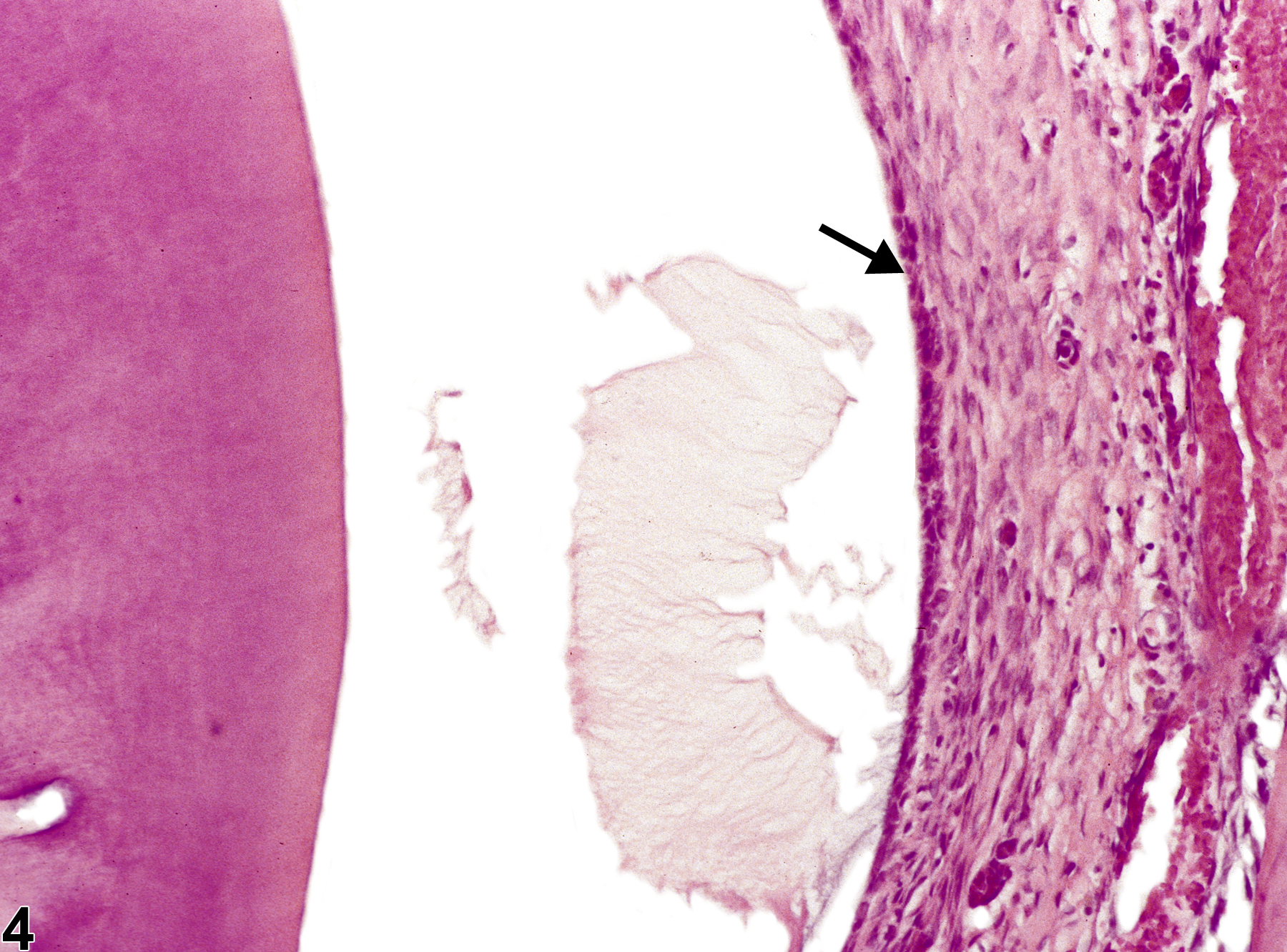
Tooth, Ameloblast - Atrophy in a male F344/N rat from a chronic study (higher magnification of Figure 2). The ameloblast layer is thin (compare with Figure 3), due to the decreased size of the ameloblasts (arrow).
Figure 4 of 4
next arrow

comment:

Ameloblasts ( Figure 1image opens in a pop-up window and Figure 3image opens in a pop-up window , arrows) are the cells responsible for enamel production. Focal atrophy of ameloblasts can lead to enamel hypoplasia, which is difficult to assess because enamel decalcification removes the enamel in tissue sections. The loss of enamel may impart a characteristic brownish discoloration on gross examination in affected teeth. Ameloblast atrophy ( Figure 2image opens in a pop-up window  and Figure 4image opens in a pop-up window , arrows) can be caused by chemical exposure, most notably fluoride toxicity. Odontoblasts can also be affected, but ameloblasts appear to be more sensitive. Atrophy is characterized by shortening of individual ameloblasts ( Figure 4image opens in a pop-up window , arrow) due to decreased amounts of cytoplasm and smaller nuclei. Atrophy can lead to squamous metaplasia of ameloblasts, characterized by loss of the columnar layer of ameloblasts with replacement by squamous epithelium (see Tooth, Ameloblast - Metaplasia, Squamous).

recommendation:

Whenever present, ameloblast atrophy should be diagnosed and graded based on the degree of cellular atrophy and the number of ameloblasts affected.

references:

Brown HR, Hardisty JF. 1990. Oral cavity, esophagus and stomach. In: Pathology of the Fischer Rat (Boorman GA, Montgomery CA, MacKenzie WF, eds). Academic Press, San Diego, CA, 9-30.
Abstract: https://www.ncbi.nlm.nih.gov/nlmcatalog/9002563

Fletcher AM, Bregman CL, Woicke J, Salcedo TW, Zidell RH, Janke HE, Fang H, Janusz WJ, Schlze GE, Mense MG. 2010. Incisor degeneration in rats induced by vascular endothelial growth factor/fibroblast growth factor receptor tyrosine kinase inhibition. Toxicol Pathol 38:267-279.
Abstract: https://www.ncbi.nlm.nih.gov/pubmed/20100840

National Toxicology Program. 1996. NTP TR-393. Toxicology and Carcinogenesis Studies of Sodium Fluoride (CAS No. 7681-49-4) in F344/N Rats and B6C3F1 Mice (Drinking Water Studies). NTP, Research Triangle Park, NC.
Abstract: https://ntp.niehs.nih.gov/go/12237