

Alimentary System
Tooth - Angiectasis
Narrative
The difference between angiectasis and hemangioma can be unclear. Hemangiomas tend to be well-circumscribed unencapsulated masses composed of tightly packed dilated vascular spaces. Each vascular space is enclosed and lined by a single layer of normal-appearing endothelial cells aligned on collagenous septa, which are usually thin, although some have broad collagenous stroma. Angiectasis does not usually present as a well-circumscribed mass, as the dilated vascular channels often course irregularly through connective tissue.
Eustis SL, Boorman GA, Harada T, Popp JA. 1990. Liver. In: Pathology of the Fischer Rat (Boorman GA, Montgomery CA, MacKenzie WF, eds). Academic Press, San Diego, CA, 71-94.
Abstract: https://www.ncbi.nlm.nih.gov/nlmcatalog/9002563Long PH, Leininger JR. 1999. Teeth. In: Pathology of the Mouse (Maronpot RR, ed). Cache River Press, St Louis, MO, 13-28.
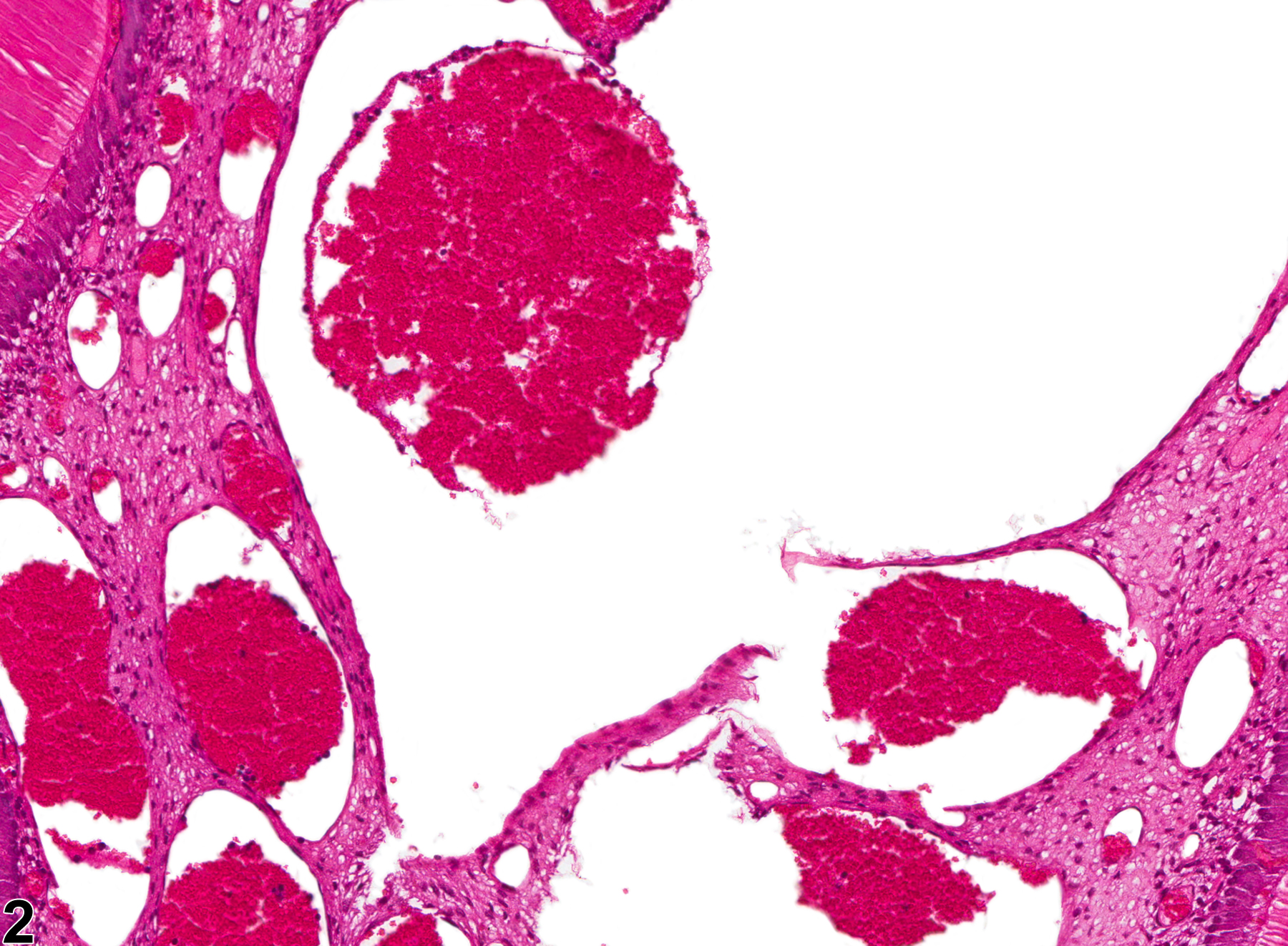
Tooth - Angiectasis in a male F344/N rat from a chronic study. There are numerous dilated blood vessels in the tooth pulp (arrow), consistent with angiectasis.
All Images

Tooth - Angiectasis in a male F344/N rat from a chronic study. There are numerous dilated blood vessels in the tooth pulp (arrow), consistent with angiectasis.

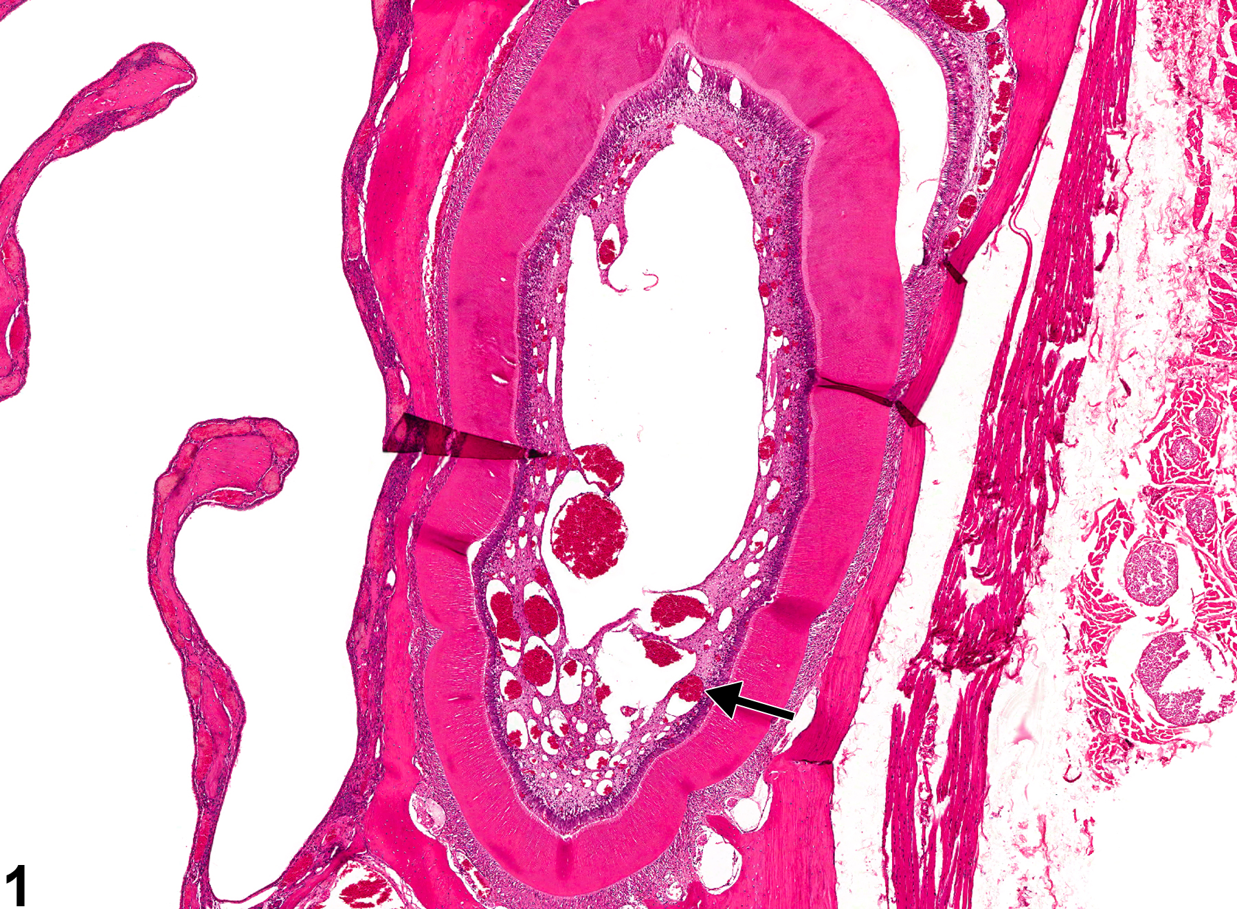
Tooth - Angiectasis in a male F344/N rat from a chronic study (higher magnification of Figure 1). There are numerous dilated blood vessels with unremarkable endothelial cells in the tooth pulp.