

Alimentary System
Tooth - Dental Dysplasia
Narrative
Dental dysplasia must be differentiated from odontoma. The term "odontoma" refers to what most authorities consider a hamartomatous malformation that resembles a neoplasm. Odontomas are characterized by proliferative redundancies of the dental organ that progress to the stage of both enamel and dentin formation. Size and cellularity/proliferative activity are criteria for differentiating odontomas from dental dysplasia. In general, dental dysplasia resembles attempted repair (reminiscent of a fracture callus with abundant dense bone/osteodentin) with some residual active dental organ tissue. The diagnosis of "odontoma" is reserved for larger, more cellular masses that appear to be proliferating beyond what one would expect for a developing tooth or attempted repair of a tooth fracture. Because of expansion, odontomas can be destructive masses.
Levy BA. 1968. Effects of experimental trauma on developing first molar teeth in rats. J Dent Res 47:323-327.
Abstract: http://jdr.sagepub.com/content/47/2/323Long PH, Leininger JR. 1999. Teeth. In: Pathology of the Mouse (Maronpot RR, ed). Cache River Press, St Louis, MO, 13-28.
Losco PE. 1995. Dental dysplasia in rats and mice. Toxicol Pathol 23:677-688.
Abstract: https://www.ncbi.nlm.nih.gov/pubmed/8772253Shafer WG, Hine MK, Levy BM. 1983. Cysts and tumors of odontogenic origin. In: Oral Pathology, 4th ed (Shafer WG, ed). Saunders, Philadelphia, PA , 258-317.
Tooth - Dental dysplasia, Bilateral in a male B6C3F1 mouse from a subchronic study. There is abnormal growth of dental tissues characteristic of dental dysplasia.
All Images

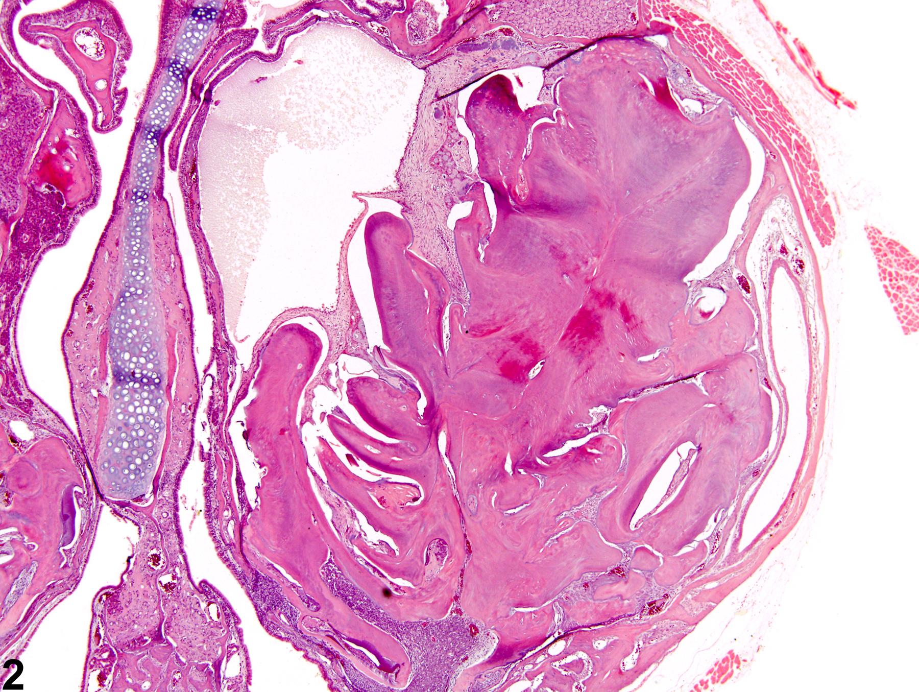
Tooth - Dental dysplasia, Bilateral in a male B6C3F1 mouse from a subchronic study. There is abnormal growth of dental tissues characteristic of dental dysplasia.

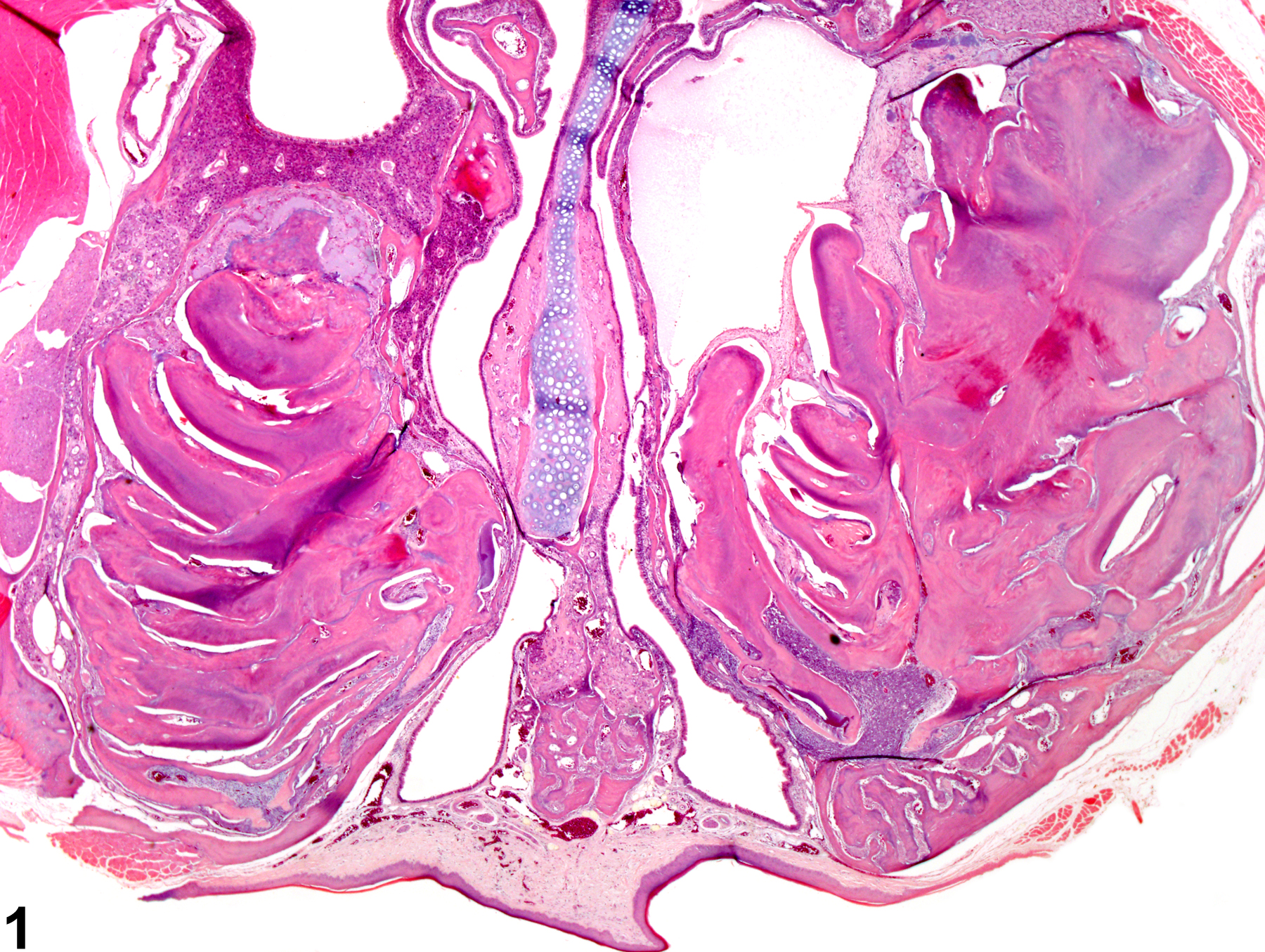
Tooth - Dental dysplasia, Bilateral in a male B6C3F1 mouse from a subchronic study (higher magnification of Figure 1). There is abnormal growth of dental tissues characteristic of dental dysplasia.

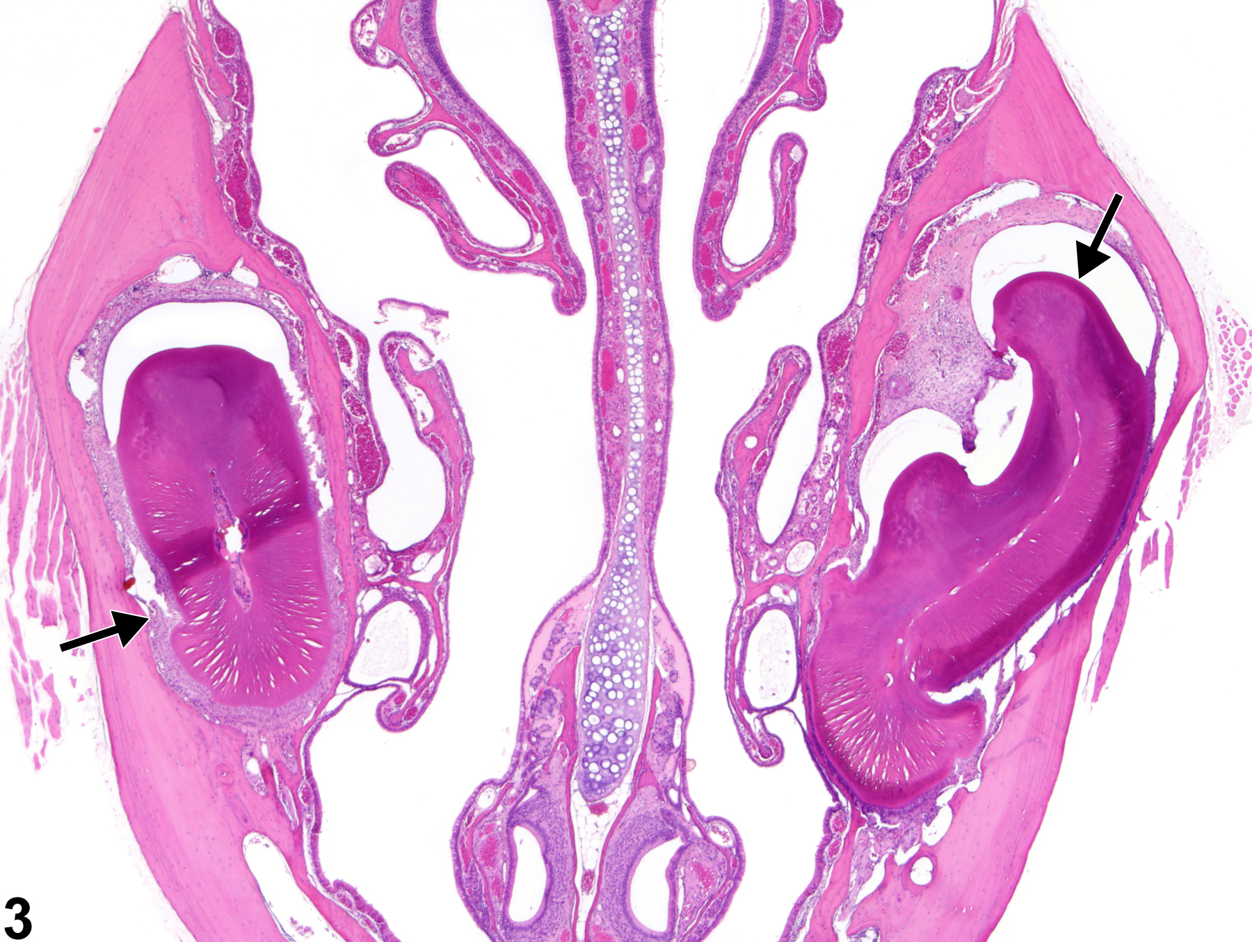
Tooth - Dental dysplasia, Bilateral in a male B6C3F1 mouse from a subchronic study. There is abnormal growth of dental tissues characteristic of dental dysplasia (arrows).

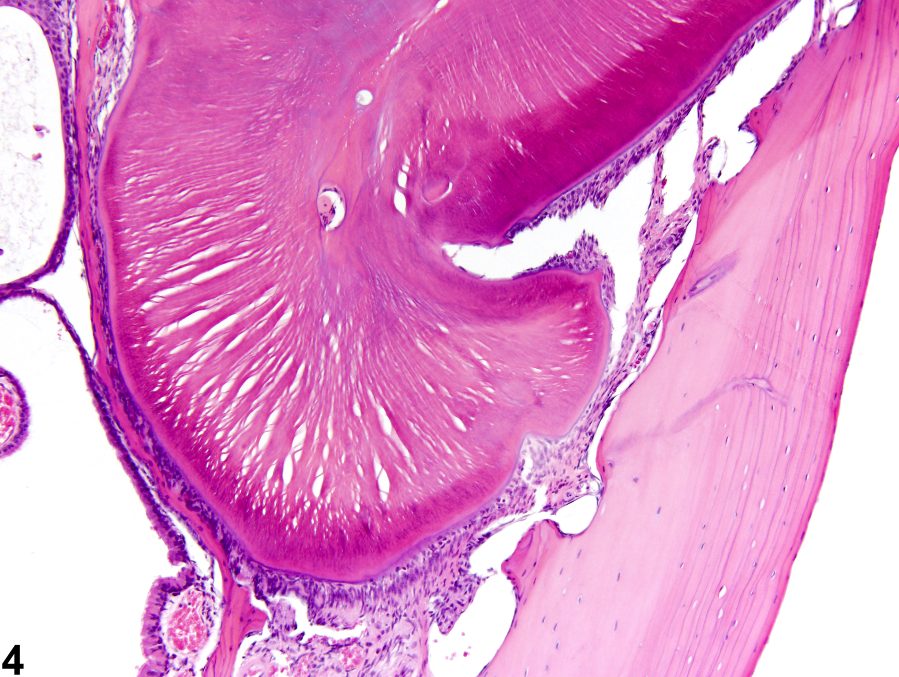
Tooth - Dental dysplasia, Bilateral in a male B6C3F1 mouse from a subchronic study (higher magnification of Figure 3). There is abnormal growth of dental tissues characteristic of dental dysplasia.