U.S. flag

An official website of the United States government

Dot gov

The .gov means it's official.
Federal government websites often end in .gov or .mil. Before sharing sensitive information, make sure you're on a federal government site.

Https

The site is secure.
The https:// ensures that you are connecting to the official website and that any information you provide is encrypted and transmitted securely.

Skip to Main Navigation
Skip to Page Content
Skip to Atlas Navigation

Tooth - Fibrosis

Image of fibrosis in the tooth from a female HSD rat in a chronic study
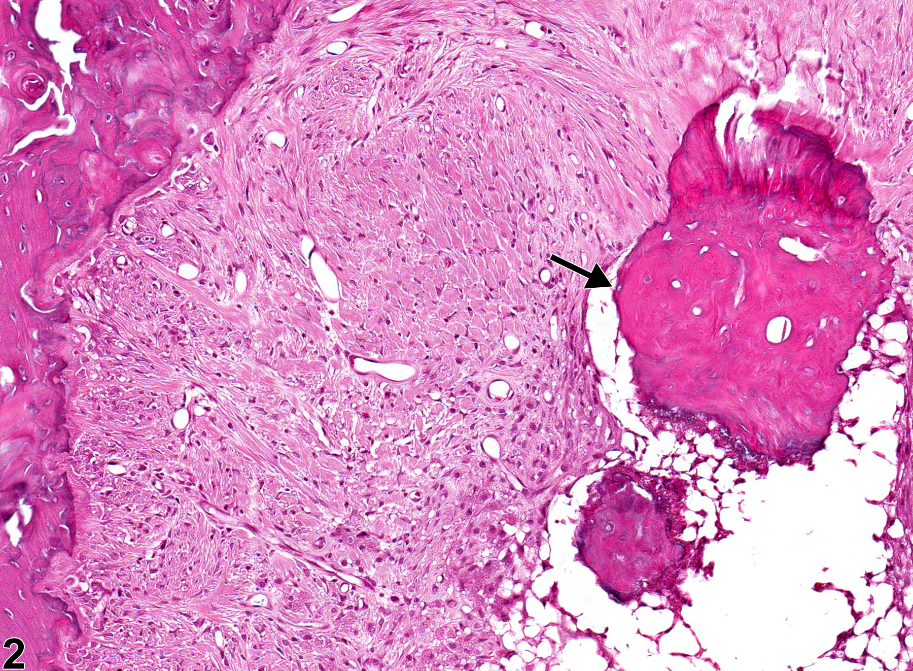
Tooth - Fibrosis in a female Harlan Sprague-Dawley rat from a chronic study. There is fibrosis within the alveolar bone and a possible sequestrum of bone or osteodentin (arrow).
Figure 1 of 4
Image of fibrosis in the tooth from a female HSD rat in a chronic study
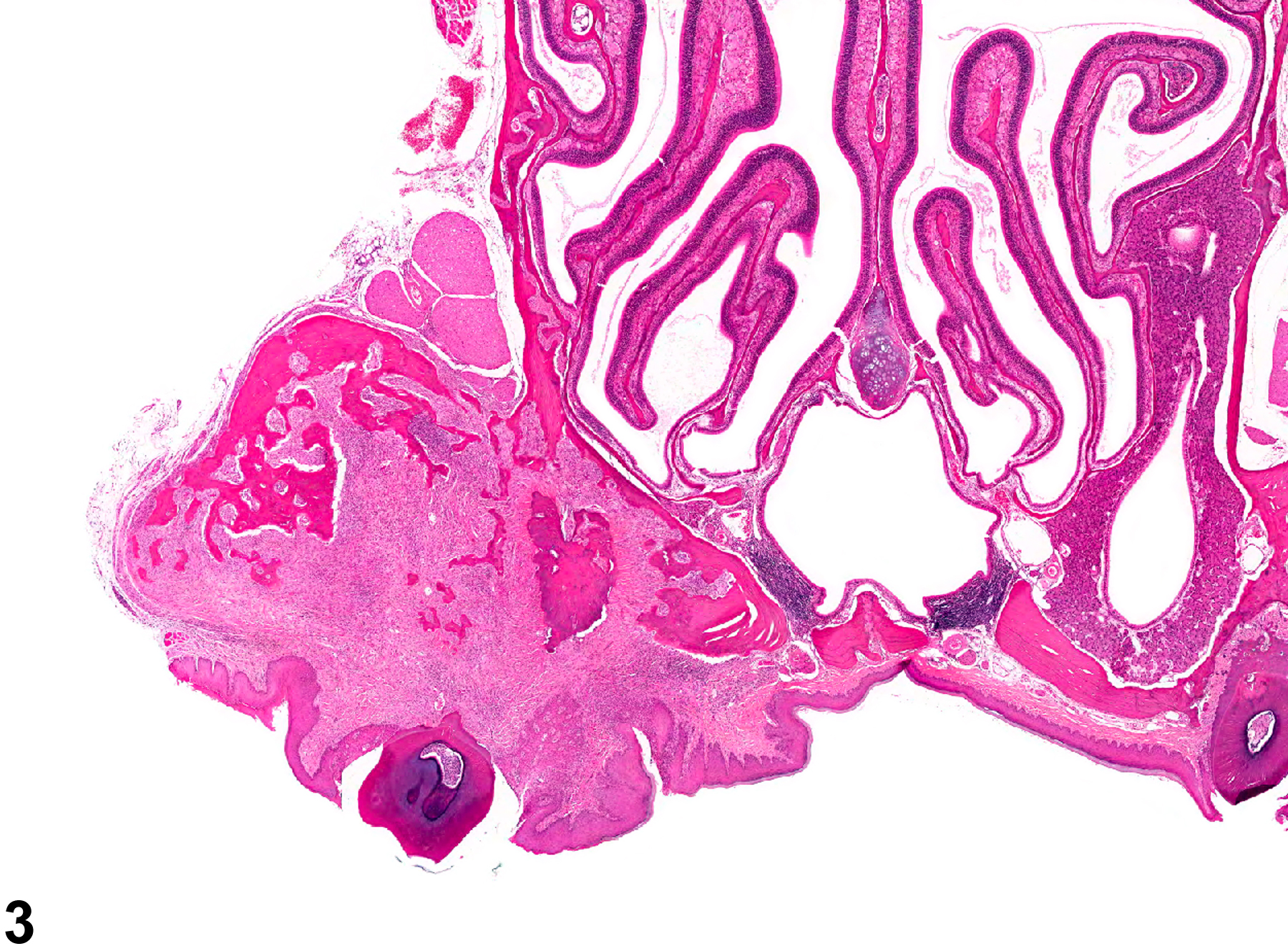
Tooth - Fibrosis in a female Harlan Sprague-Dawley rat from a chronic study (higher magnification of Figure 1). There is fibrosis within the alveolar bone and a possible sequestrum of bone or osteodentin (arrow).
Figure 2 of 4
Image of fibrosis in the tooth from a female B6C3F1 mouse in a chronic study
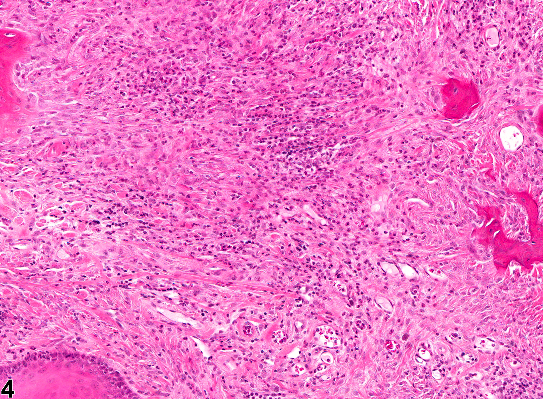
Tooth - Fibrosis in a female B6C3F1 mouse from a chronic study. Fibrosis has replaced much of the alveolar bone.
Figure 3 of 4
Image of fibrosis in the tooth from a female B6C3F1 mouse in a chronic study
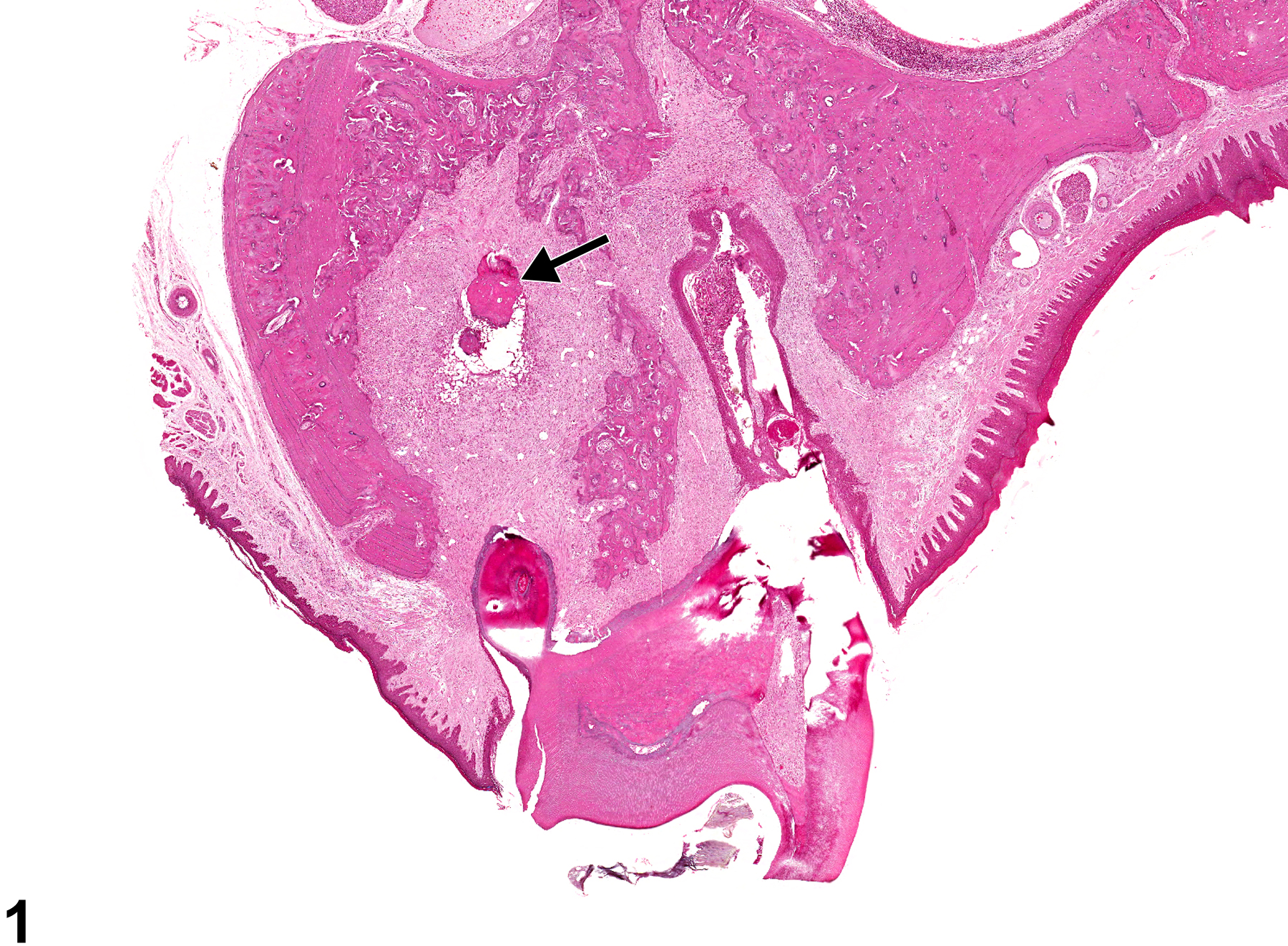
Tooth - Fibrosis in a female B6C3F1 mouse from a chronic study (higher magnification of Figure 3). Fibrosis with inflammatory cells has replaced much of the alveolar bone.
Figure 4 of 4
next arrow

comment:

Fibrosis within the alveolus ( Figure 1image opens in a pop-up window , Figure 2image opens in a pop-up window , Figure 3image opens in a pop-up window , and Figure 4image opens in a pop-up window ) may be the result of resorption affecting the incisor or molar roots. Resorption may be initiated by malocclusion, infection, or trauma (including iatrogenic fracture). Resorption may progress to complete loss of the tooth and replacement with fibrous connective tissue and/or bone. Inflammation may accompany the fibrosis ( Figure 3image opens in a pop-up window and Figure 4image opens in a pop-up window ).

recommendation:

Fibrosis should be diagnosed and graded when it is prominent in a lesion or if it has progressed to the point that fibrosis is the only lesion present. If fibrosis is a minor component of inflammation, it is not necessary to diagnose it separately, though it should be described in the pathology narrative.

references:

Long PH, Leininger JR. 1999. Teeth. In: Pathology of the Mouse (Maronpot RR, ed). Cache River Press, St Louis, MO, 13-28.
Abstract: http://www.cacheriverpress.com/books/pathmouse.htm