

Alimentary System
Tooth, Incisor - Degeneration
Narrative
Long PH, Herbert RA, Nyska A. 2004. Hexachlorobenzene-induced incisor degeneration in Sprague-Dawley rats. Toxicol Pathol 32:35-40.
Abstract: https://www.ncbi.nlm.nih.gov/pubmed/14713546Ten Cate AR. 1998. Dentin-pulp complex. In: Oral Histology: Development, Structure, and Function, 5th ed. Mosby-Year Book, St Louis, MO, 150-196.
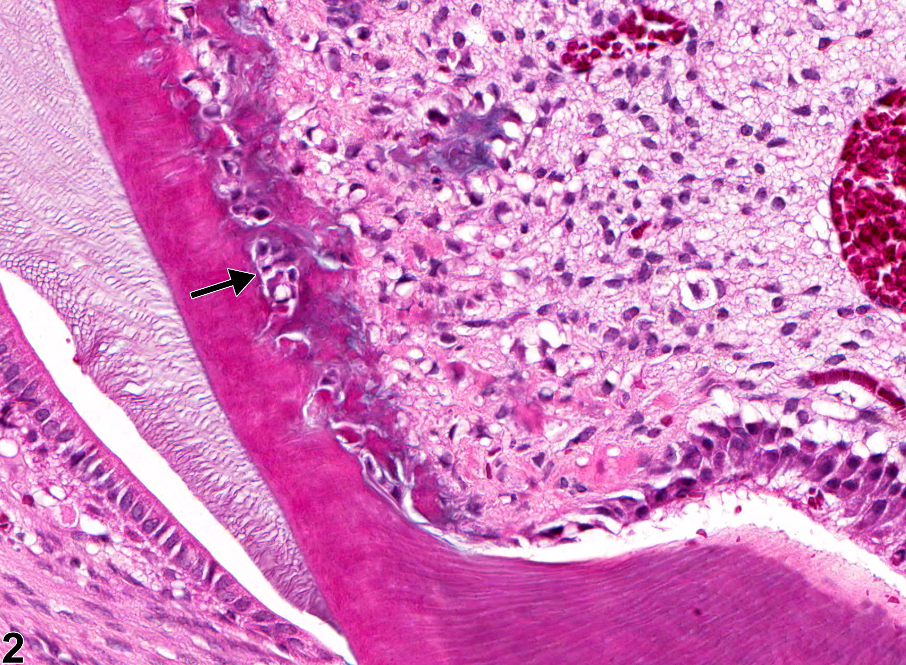
Tooth, Incisor - Degeneration in a female Harlan Sprague-Dawley rat from a subchronic study. Odontoblast degeneration and dentin niche formation (arrows) are present.
All Images

Tooth, Incisor - Degeneration in a female Harlan Sprague-Dawley rat from a subchronic study. Odontoblast degeneration and dentin niche formation (arrows) are present.

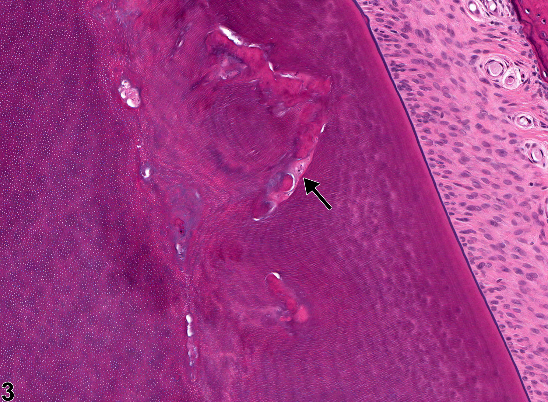
Tooth, Incisor - Degeneration in a female Harlan Sprague-Dawley rat from a subchronic study (higher magnification of Figure 1). There is thinning of the dentin layer and loss of and degeneration of odontoblasts (arrow) in a dentin niche.

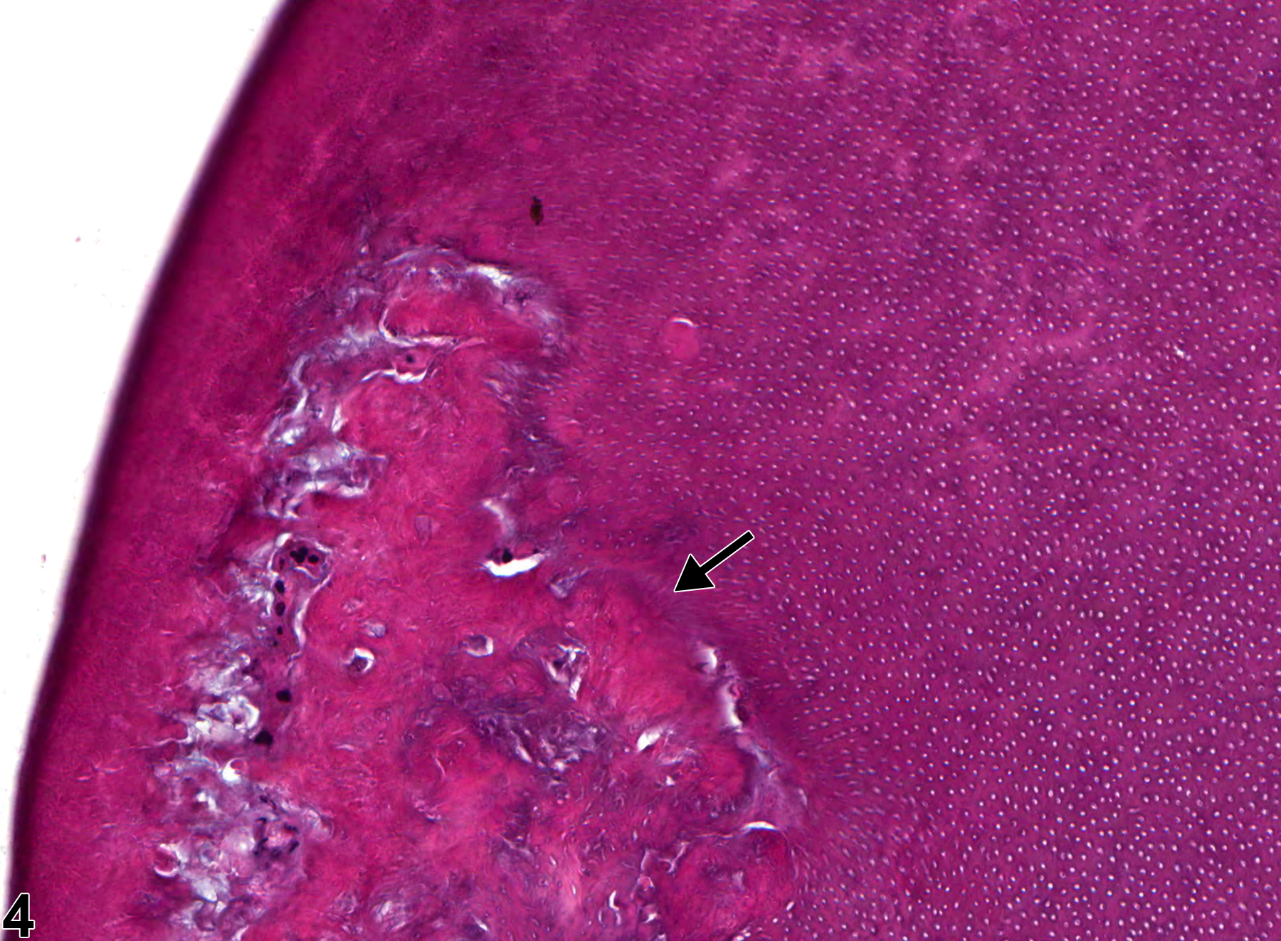
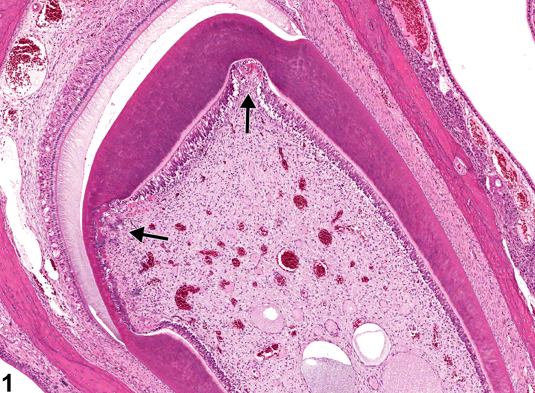
Tooth, Incisor - Degeneration in a female Harlan Sprague-Dawley rat from a subchronic study. Abnormal dentin and osteodentin production (arrow) are present.

Tooth, Incisor - Degeneration in a female Harlan Sprague-Dawley rat from a subchronic study. Abnormal dentin and osteodentin production (arrow) are present.