

Alimentary System
Tooth - Periodontal Pocket
Narrative
Differential diagnoses for periodontal pocket include squamous cyst and cystic keratinizing hyperplasia. Cystic keratinizing hyperplasia is thought to be on a continuum from hyperplasia to squamous cell carcinoma. In comparison, periodontal pockets tend to be unilocular structures lined by normal squamous epithelium. Squamous cysts are similar to periodontal pockets morphologically, but they are not necessarily located adjacent to a tooth and do not communicate with the oral mucosa. Ideally, the communication to the oral cavity can be seen in a periodontal pocket lesion, but the opening may not be apparent because of the plane of section.
Bertram TA, Markovits JE, Juliana MM. 1996. Non-proliferative lesions of the alimentary canal in rats GI-1. In Guides for Toxicologic Pathology. STP/ARP/AFIP, Washington, DC, 1-16.
Full Text: https://www.toxpath.org/docs/SSNDC/GINonproliferativeRat.pdfNational Toxicology Program. 2010. NTP TR-558. Toxicology and Carcinogenesis Studies of 3,3’,4,4’-Tetrachloroazobenzene (TCAB) (CAS No. 14047-09-7) in Harlan Sprague Dawley Rats and B6C3F1 Mice (Gavage Studies). NTP, Research Triangle Park, NC.
Abstract: https://ntp.niehs.nih.gov/go/33564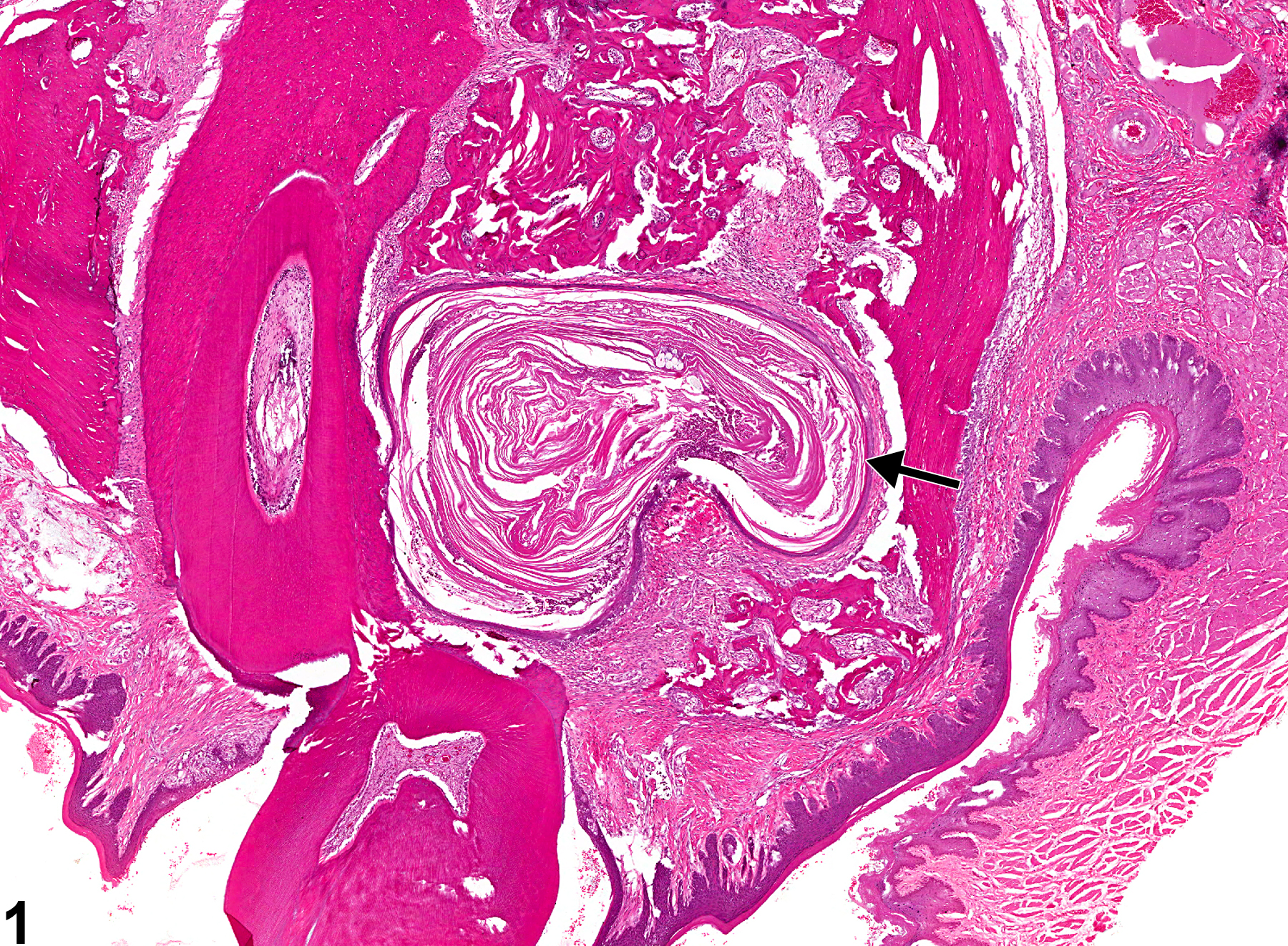
Tooth - Periodontal pocket in a male F344/N rat from a chronic study. The opening of the pocket (arrow) to the oral cavity is not apparent because of the plane of section.
All Images

Tooth - Periodontal pocket in a male F344/N rat from a chronic study. The opening of the pocket (arrow) to the oral cavity is not apparent because of the plane of section.

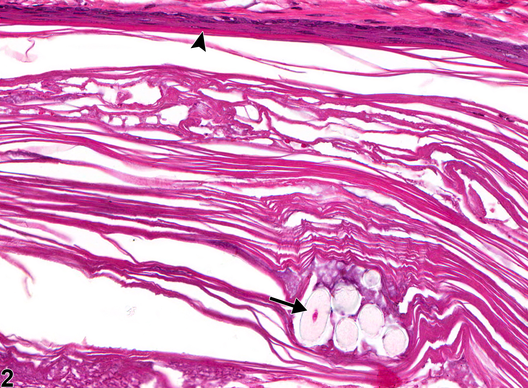
Tooth - Periodontal pocket in a male F344/N rat from a chronic study (higher magnification of Figure 1). The periodontal pocket is lined by squamous epithelium (arrowhead and contains hair shafts (arrow).

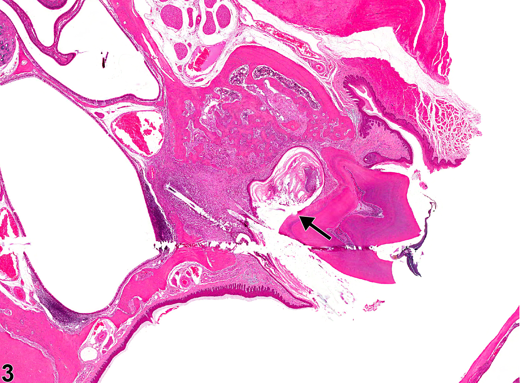
Tooth - Periodontal pocket in a male F344/N rat from a chronic study. The opening of the pocket (arrow) to the oral cavity is adjacent to the tooth.

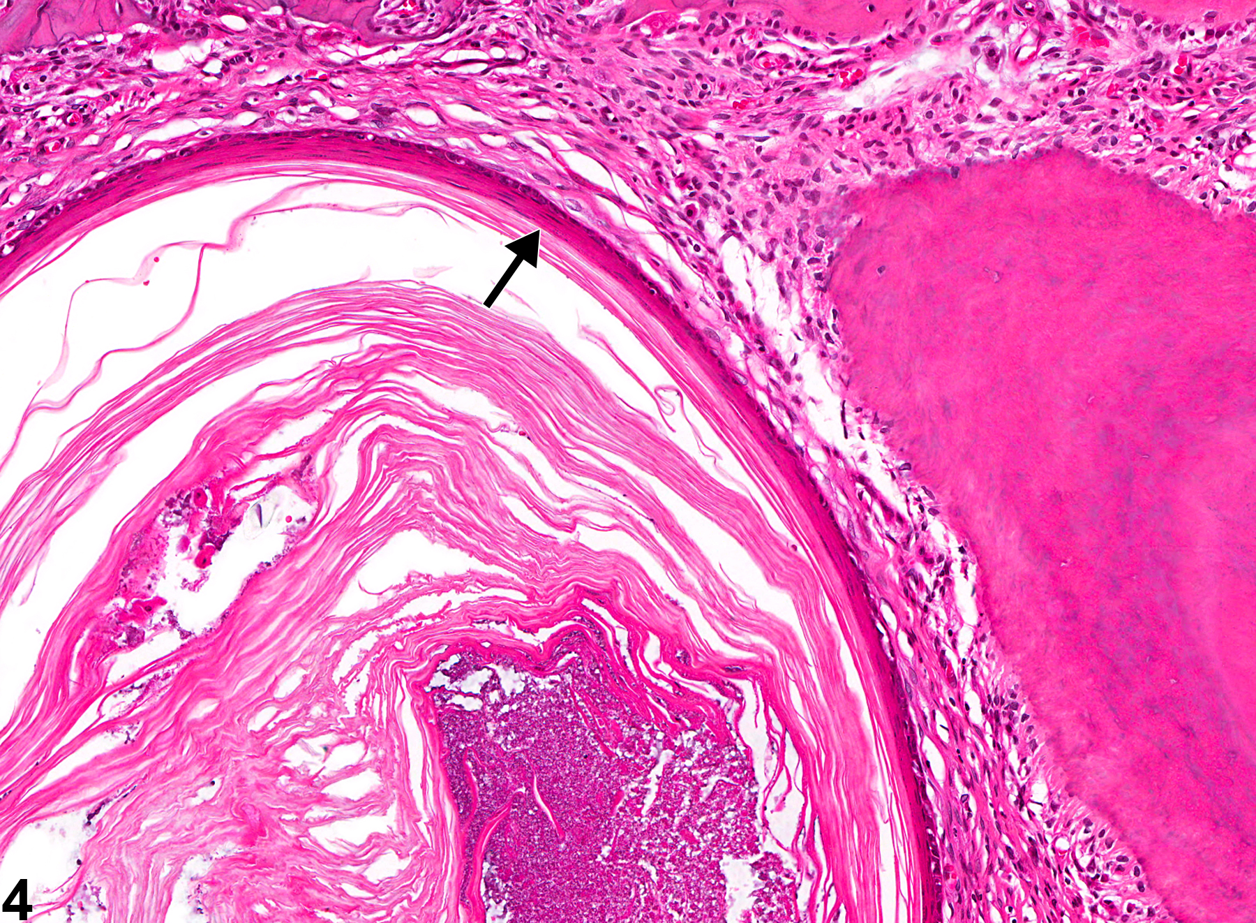
Tooth - Periodontal pocket in a male F344/N rat from a chronic study (higher magnification of Figure 3). The pocket is lined by keratinizing squamous epithelium (arrow) and is surrounded by fibrosis and chronic inflammation.

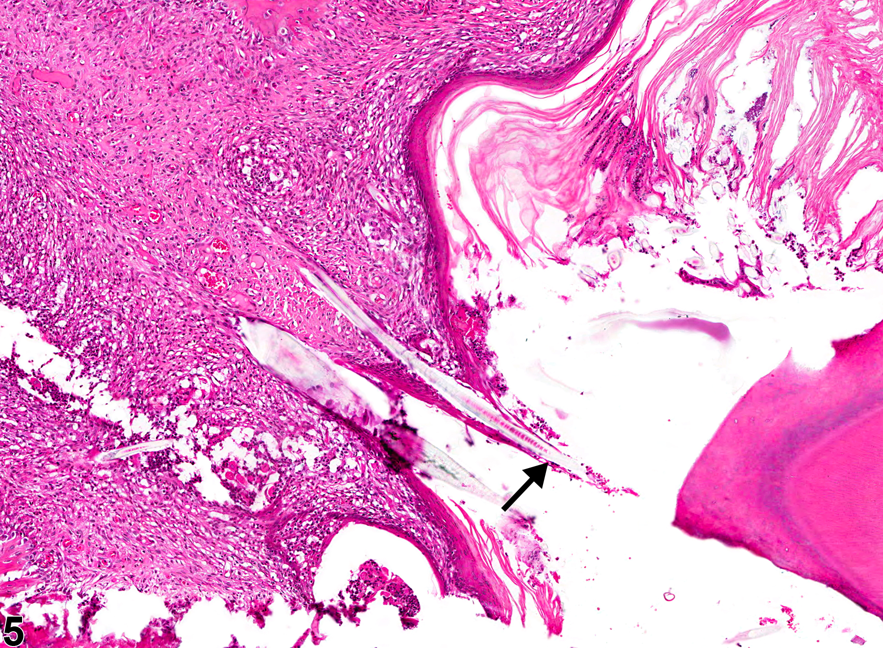
Tooth - Periodontal pocket in a male F344/N rat from a chronic study (higher magnification of Figure 3). Hair shafts (arrow) are embedded in the periodontal tissue at the opening of the pocket.

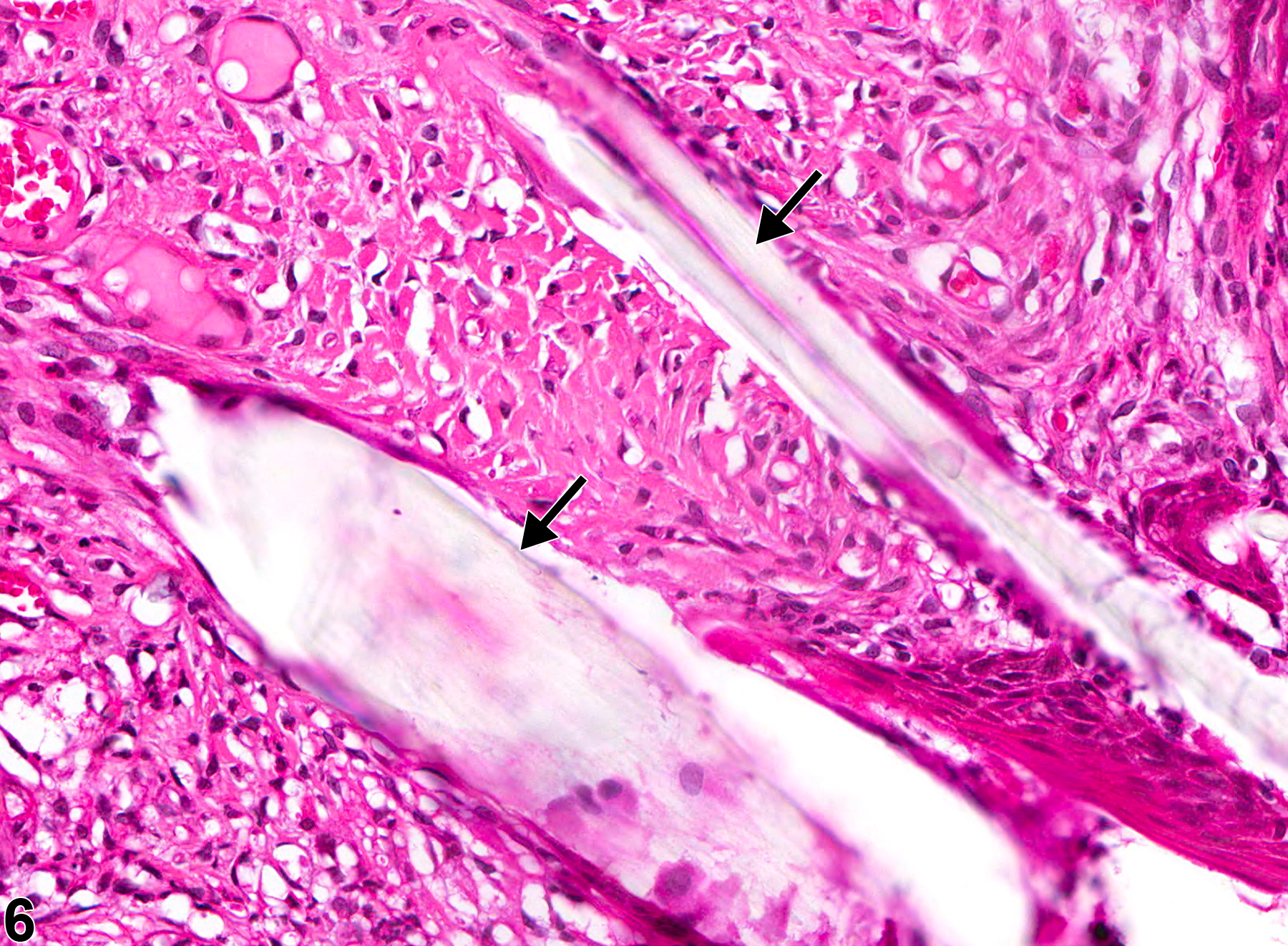
Tooth - Periodontal pocket in a male F344/N rat from a chronic study (higher magnification of Figure 3). Hair shafts (arrows) are embedded in the periodontal tissue.

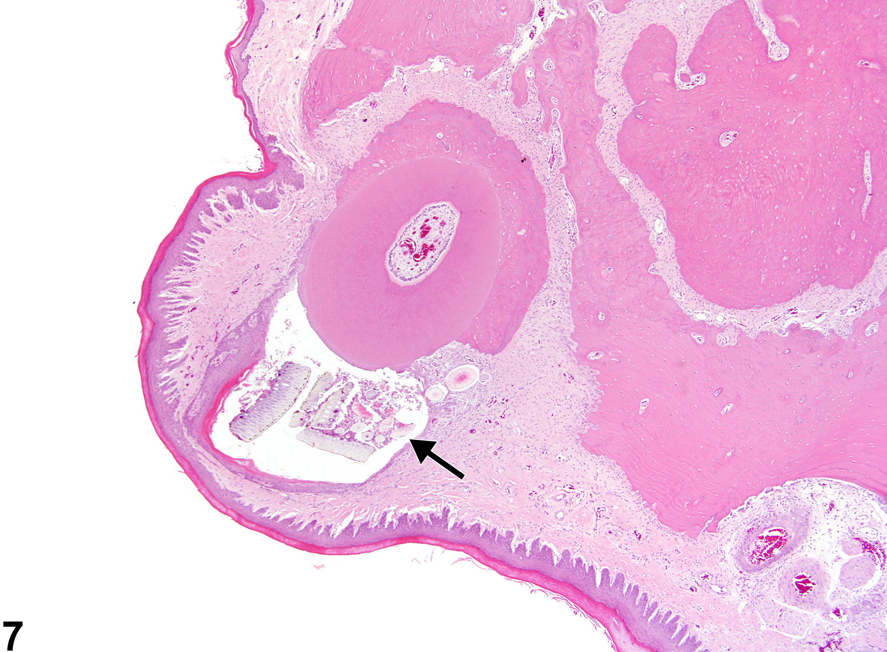
Tooth - Periodontal pocket in a male F344/N rat from a chronic study. The opening of the pocket (arrow) to the oral cavity is not apparent because of the plane of section.

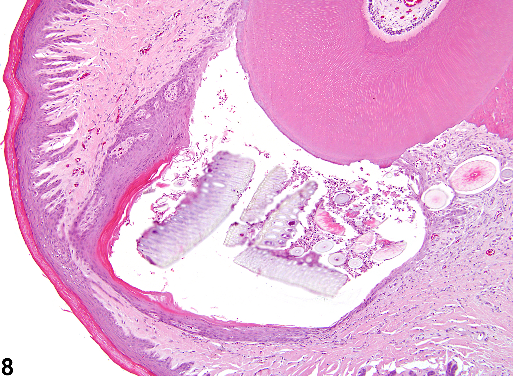
Tooth - Periodontal pocket in a male F344/N rat from a chronic study (higher magnification of Figure 7). The periodontal pocket is lined by hyperplastic, keratinizing squamous epithelium and contains feed material.