

Cardiovascular System
Blood Vessel
Narrative
Blood vessels may be one of the most important tissues examined due to their ubiquitous and extensive presence in every organ. There are three main types of blood vessels: arteries, veins, and capillaries. Arteries and arterioles carry blood away from the heart (Figure 1, Figure 2, Figure 3). Veins and venules carry blood to the heart. An exception to this generalization are the portal veins, which carry blood from one organ to another (e.g., the hepatic portal vein, which carries blood from the gut to the liver). Capillaries are the smallest vessels and are the site of exchange of oxygen, nutrients, and waste products between the tissues and the blood.
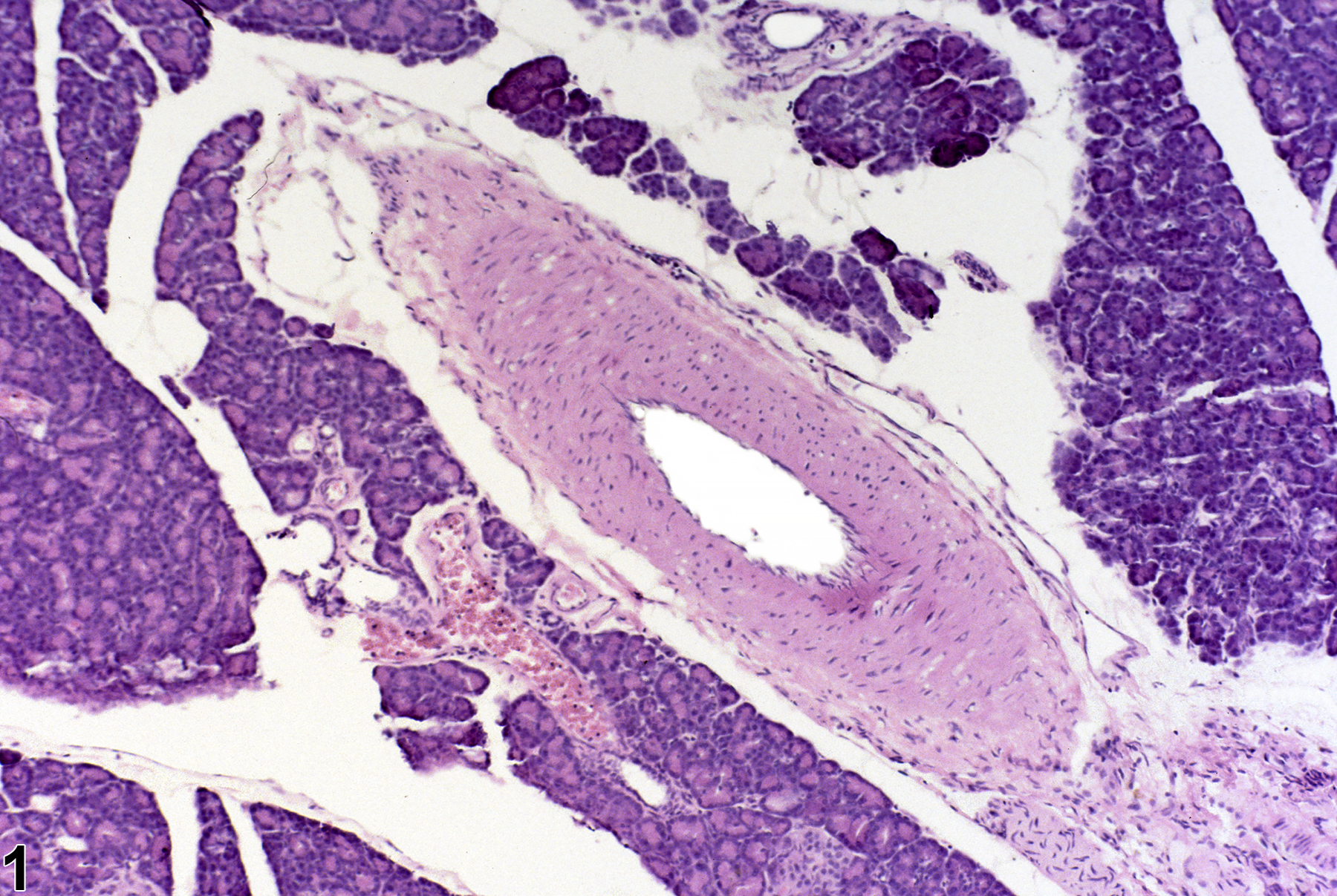
Figure 1. Normal artery within the pancreas in a male F344/N rat from a chronic study. The smooth muscle layer is the thickest layer.
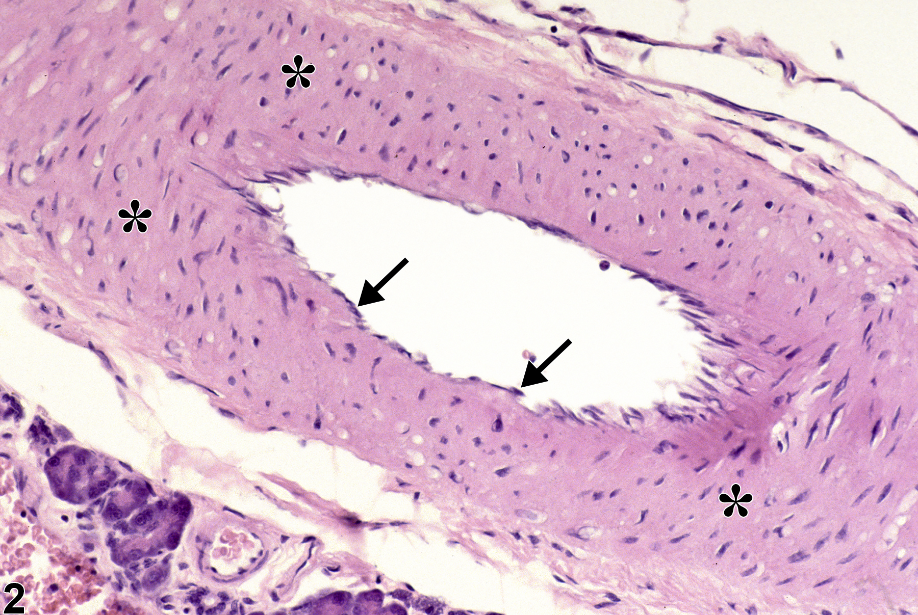
Figure 2. Normal tunica media (asterisks) and tunica intima (arrows) of an artery within the pancreas in a male F344/N rat from a chronic study (higher magnification of Figure 1).
Arteries and veins have walls composed of three layers: the tunica intima, the tunica media, and the tunica adventitia. The tunica intima is composed of endothelial cells on a basement membrane and a subendothelial layer of collagen and elastic fibers. The tunica media consists of smooth muscle cells, elastic fibers, and collagen. The tunica adventitia is composed of collagen and elastic fibers. The relative amounts of each of these components vary depending on the type of vessel. The tunica intima and tunica media of elastic arteries are generally thicker than those of the other types, and the tunica media contains elastic fibers. Capillaries differ in that they have a very thin tunica adventitia and their endothelial cells are fenestrated to increase their permeability. Capillaries in the brain lack a tunica adventitia.
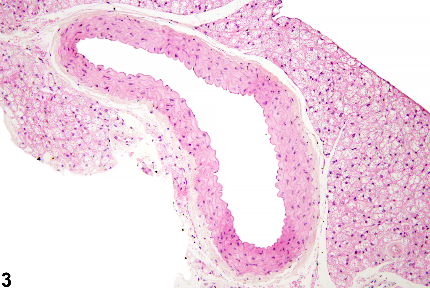
Figure 3. Normal aorta in a male B6C3F1/N mouse from a chronic study.
Because of their distribution, blood vessels may be the target of test-article-induced lesions in any organ system examined. In addition, spontaneous lesions occur quite commonly in various strains of rats and mice, and differentiation of these lesions from treatment-related lesions can be challenging. Polyarteritis nodosa is a spontaneous disease of rodents that may be exacerbated by chemical administration and is a confounding factor in the interpretation of toxicologic findings in blood vessels.
In NTP studies, changes in blood vessels should be documented in the organ in which they are found and the type of blood vessel identified (e.g., artery or vein). The aorta is considered its own tissue (organ), and lesions in the aorta are recorded as such.
Elwell MR, Mahler JF. 1999. Heart, blood vessels, and lymphatic vessels. In: Pathology of the Mouse: Reference and Atlas (Maronpot RR, Boorman GA, Gaul BW, eds). Cache River Press, Vienna, IL, 361-380.
Mitsumori K. 1990. Blood and lymphatic vessels. In: Pathology of the Fischer Rat: Reference and Atlas (Boorman GA, Eustis SL, Elwell MR, Montgomery CA, MacKenzie WF, eds). Academic Press, San Diego, CA, 473-483.