U.S. flag

An official website of the United States government

Dot gov

The .gov means it's official.
Federal government websites often end in .gov or .mil. Before sharing sensitive information, make sure you're on a federal government site.

Https

The site is secure.
The https:// ensures that you are connecting to the official website and that any information you provide is encrypted and transmitted securely.

Skip to Main Navigation
Skip to Page Content
Skip to Atlas Navigation

Heart - Amyloid

Image of amyloid in the heart from a male Swiss Webster mouse in a chronic study
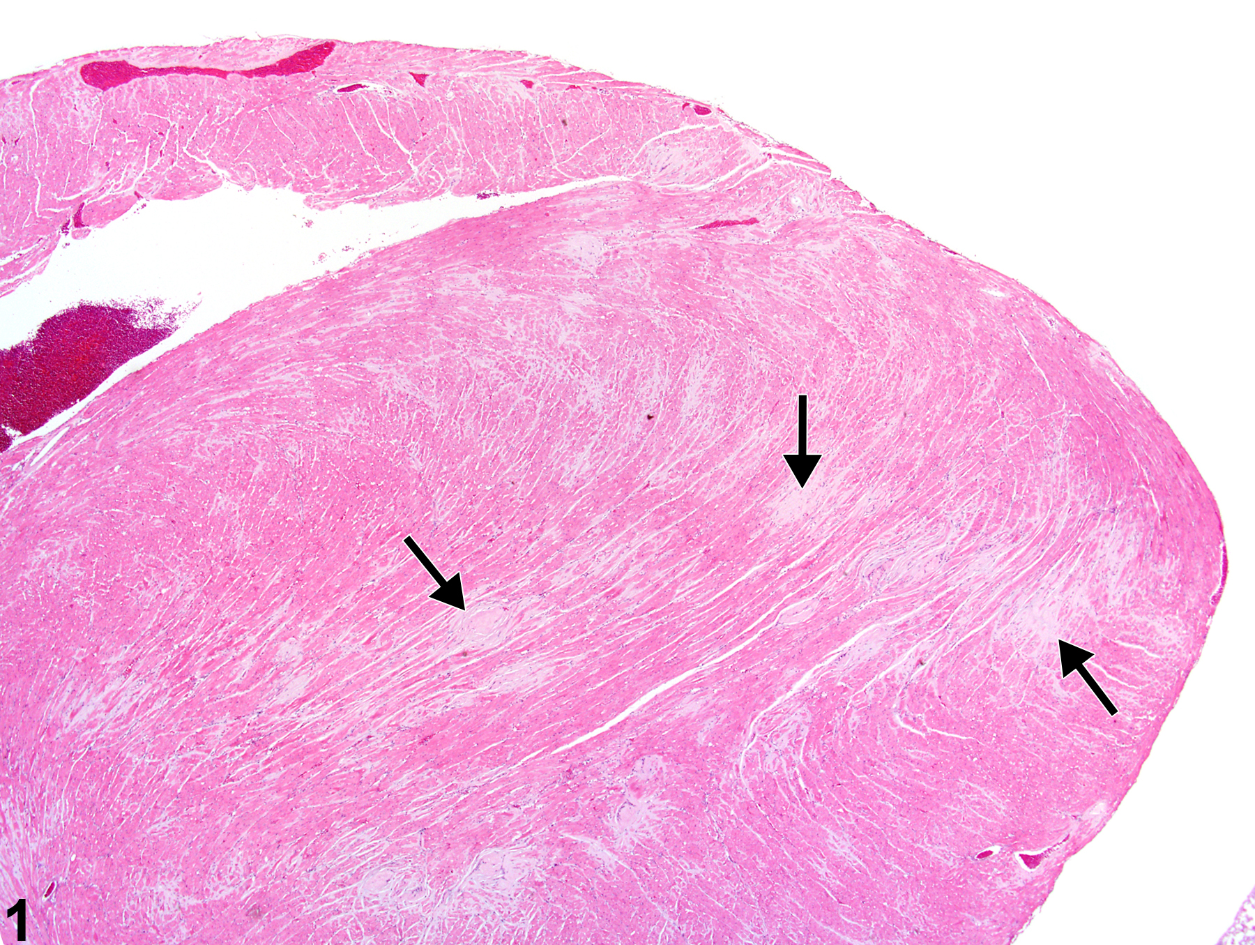
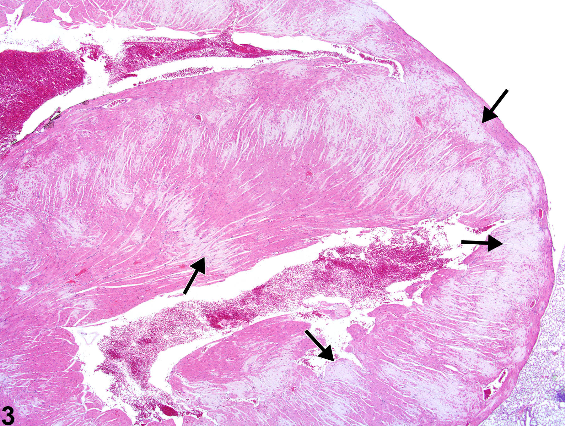
Heart - Amyloid in a male Swiss Webster mouse from a chronic study. Multiple amyloid plaques (arrows) are present in the myocardium.
Figure 1 of 4
Image of amyloid in the heart from a male Swiss Webster mouse in a chronic study
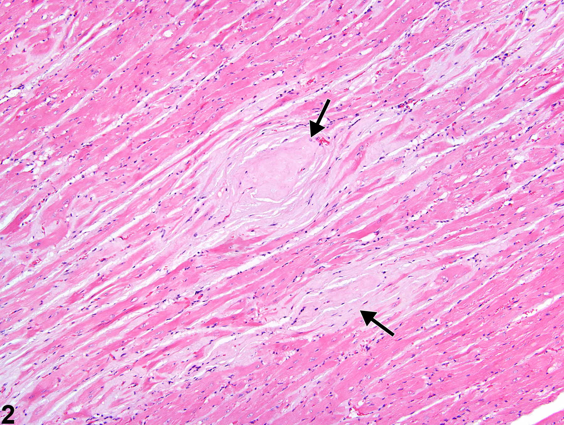
Heart - Amyloid in a male Swiss Webster mouse from a chronic study (higher magnification Figure 1). The amyloid (arrows) is composed of acellular, eosinophilic material.
Figure 2 of 4
Image of amyloid in the heart from a male Swiss Webster mouse in a chronic study
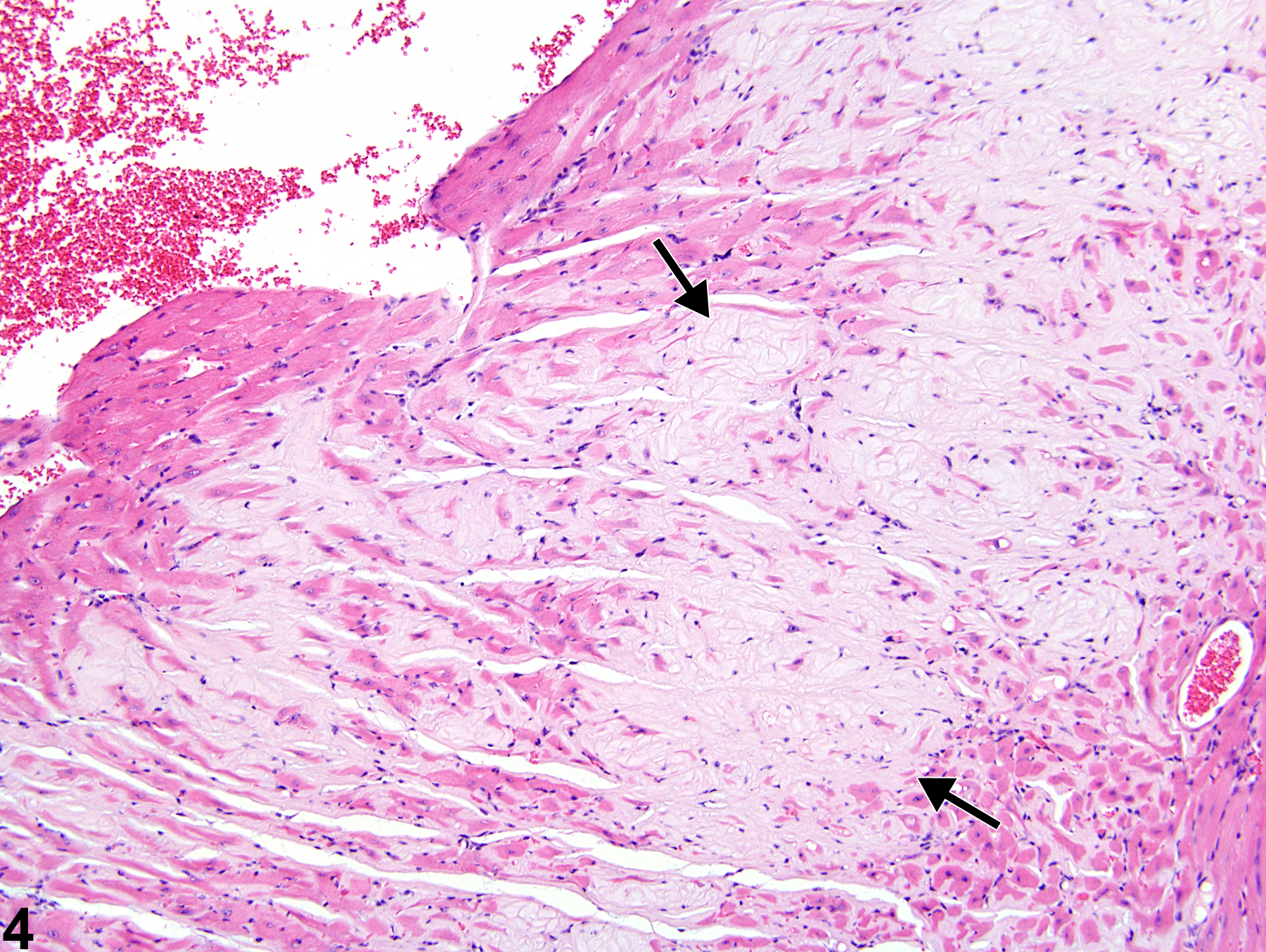
Heart - Amyloid in a male Swiss Webster mouse from a chronic study. Multiple pale areas contain amyloid (arrows).
Figure 3 of 4
Image of amyloid in the heart from a male Swiss Webster mouse in a chronic study
Heart - Amyloid in a male Swiss Webster mouse from a chronic study (higher magnification of Figure 3). Coalescing areas of pale, eosinophilic, homogeneous amyloid are present (arrows).
Figure 4 of 4
next arrow

comment:

Amyloid (amyloidosis) is a systemic disease that is rare in B6C3F1, BALB/c, and C3H/HeJ mice and in rats but is common in CD-1, A, Swiss Webster, SJL, and C57BL/6 mice and can be a cause of death. The incidence can be increased in association with fighting among group-housed males and with ectoparasitism. The Swiss Webster mouse strain has been reported to exhibit a high incidence of cardiac amyloidosis, which is related to chronic dermatitis. Amyloid appears as an amorphous, eosinophilic, hyaline, extracellular substance ( Figure 1image opens in a pop-up window , Figure 2image opens in a pop-up window , Figure 3image opens in a pop-up window , and Figure 4image opens in a pop-up window ) that, with progressive accumulation, results in pressure atrophy of adjacent cells and tissue, leading to functional impairment. Congo red stains amyloid orange to orange red and under polarized light imparts a light green, so-called apple green, fluorescence.

recommendation:

Heart - Amyloid should be diagnosed and graded based on the extent of deposition. Degeneration of cells secondary to amyloid deposition should not be diagnosed separately unless warranted by severity. If amyloid is present within the wall of a blood vessel, then the type of blood vessel (e.g., arerty, vein) should be included as a site modifier. If amyloid occurs in multiple sites,these may be listed in the narrative. The modifier "multiple" should not be used in the diagnosis. Rather, the total area affected by amyloid should be reflected in the severity grade.

references:

Bucher JR, Shackelford CC, Hasemen JK, Johnson JD, Kurtz PJ, Persing RL. 1994. Carcinogenicity studies of oxazepam in mice. Fund Appl Toxicol 23:280-297.
Abstract: https://www.ncbi.nlm.nih.gov/pubmed/7982536

Elwell MR, Mahler JF. 1999. Heart, blood vessels, and lymphatic vessels. In: Pathology of the Mouse: Reference and Atlas (Maronpot RR, Boorman GA, Gaul BW, eds). Cache River Press, Vienna, IL, 361-380.

Gruys E, Tooten PC, Kuijpers MH. 1996. Lung, ileum and heart are predilection sites for AApoAII amyloid deposition in CD-1 Swiss mice used for toxicity studies. Pulmonary amyloid indicates AApoAII. Lab Anim 30:28-34.
Abstract: https://www.ncbi.nlm.nih.gov/pubmed/8709570

Maita K, Hirano M, Harada T, Mitsumori K, Yoshida A, Takahashi K, Nakashima N, Kitazawa T, Enomoto A, Inui K, Shirasu Y. 1988. Mortality, major cause of moribundity, and spontaneous tumors in CD-1 mice. Toxicol Pathol 16:340-349.
Abstract: https://www.ncbi.nlm.nih.gov/pubmed/3194656

Yoshizawa K, Kissling GE, Johnson JA, Clayton NP, Flagler ND, Nyska A. 2005. Chemical-induced atrial thrombosis in NTP rodent studies: Potential mechanisms and literature review. Toxicol Pathol 33:517-532.
Full Text: http://tpx.sagepub.com/content/33/5/517.full