

Cardiovascular System
Heart - Atrophy
Narrative
Campbell SE, Korecky B, Rakusan K. 1991. Remodeling of myocyte dimensions in hypertrophic and atrophic rat hearts. Circ Res 68:984-996.
Abstract: https://www.ncbi.nlm.nih.gov/pubmed/1826234Elwell MR, Mahler JF. 1999. Heart, blood vessels, and lymphatics. In: Pathology of the Mouse: Reference and Atlas (Maronpot RR, Boorman GA, Gaul BW, eds). Cache River Press, Vienna, IL, 361- 380.
Greaves P. 2007. Cardiovascular system. In: Histopathology of Preclinical Toxicity Studies, 3rd ed (Greaves P, ed). Academic Press, New York, 270-333.
Ito T, Kimura Y, Uozumi Y, Takai M, Muraoka S, Matsuda T, Ueki K, Yoshiyama M, Ikawa M, Okabe M, Schaffer SW, Fujio Y, Azuma J. 2008. Taurine depletion caused by knocking out the taurine transporter gene leads to cardiomyopathy with cardiac atrophy. J Mol Cell Cardiol 44:927-937.
Abstract: https://www.ncbi.nlm.nih.gov/pubmed/18407290Jokinen MP, Lieuallen WG, Johnson CL, Dunnick J, Nyska A. 2005. Characterization of spontaneous and chemically induced cardiac lesions in rodent model systems: The National Toxicology Program experience. Cardiovasc Toxicol 5:227-244.
Abstract: https://www.ncbi.nlm.nih.gov/pubmed/16046796McKenzie WF, Alison R. 1990. Heart. In: Pathology of the Fischer Rat: Reference and Atlas (Boorman GA, Eustis SL, Elwell MR, Montgomery CA, MacKenzie WF, eds). Academic Press, San Diego, CA, 461-472.
Percy DH, Barthold SW. 2007. Pathology of Laboratory Rodents and Rabbits, 3rd ed. Blackwell, Ames, IA, 152-153.
Razeghi P, Baskin KK, Sharma S, Young ME, Stepkowski S, Essop MF, Taegtmeyer H. 2006. Atrophy, hypertrophy, and hypoxemia induce transcriptional regulators of the ubiquitin proteasome system in the rat heart. Biochem Biophys Res Commun 342:361-364.
Abstract: https://www.ncbi.nlm.nih.gov/pubmed/16483544Razeghi P, Volpini KC, Wang ME, Youker KA, Stepkowski S, Taegtmeyer H. 2007. Mechanical unloading of the heart activates the calpain system. J Mol Cell Cardiol 42:449-452.
Abstract: https://www.ncbi.nlm.nih.gov/pubmed/17027024Van Vleet JF, Ferrans VJ. 2007. Cardiovascular system. In: Pathologic Basis of Veterinary Disease, 4th ed (McGavin MD, Zachary JF, eds). Mosby, St. Louis, MO, 575-584.
Van Vleet JF, Ferrans VJ, Herman E. 2010. Cardiovascular and skeletal muscle systems. In: Handbook of Toxicologic Pathology, 2nd ed (Haschek WM, Rousseaux CG, Wallig MA, eds). Academic Press, New York, 335-346.
Zhu W, Shou W, Payne RM, Caldwell R, Field LJ. 2008. A mouse model for juvenile doxorubicin-induced cardiac dysfunction. Pediatr Res 64:488-494.
Abstract: https://www.ncbi.nlm.nih.gov/pubmed/18614963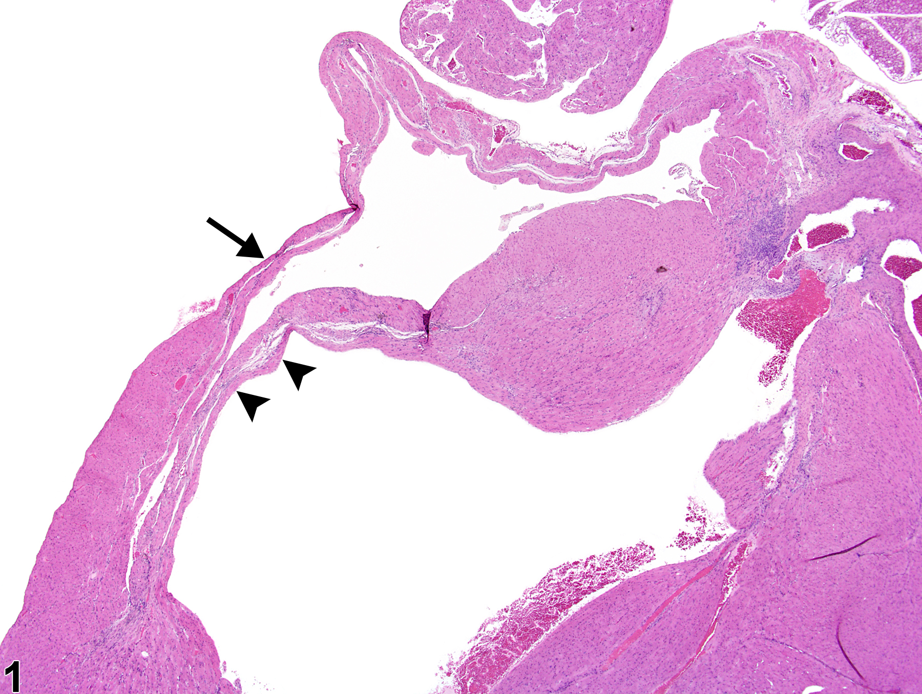
Heart - Atrophy in a male B6C3F1/N mouse from a chronic study. Thinning of the right ventricular wall (arrow) and interventricular septum (arrowheads) is present.
All Images

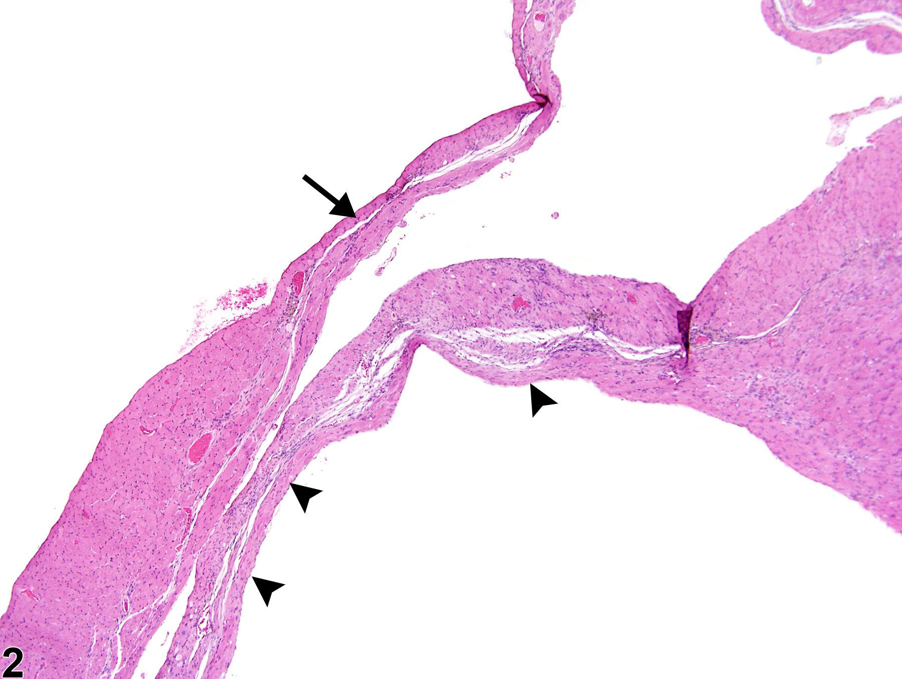
Heart - Atrophy in a male B6C3F1/N mouse from a chronic study. Thinning of the right ventricular wall (arrow) and interventricular septum (arrowheads) is present.

Heart - Atrophy in a male B6C3F1/N mouse from a chronic study (higher magnification of Figure 1). Atrophy is present in the right ventricular wall (arrow) and interventricular septum (arrowheads).