U.S. flag

An official website of the United States government

Dot gov

The .gov means it's official.
Federal government websites often end in .gov or .mil. Before sharing sensitive information, make sure you're on a federal government site.

Https

The site is secure.
The https:// ensures that you are connecting to the official website and that any information you provide is encrypted and transmitted securely.

Skip to Main Navigation
Skip to Page Content
Skip to Atlas Navigation

Heart - Hemorrhage

Image of hemorrhage in the heart from a male F344/N rat in a acute study
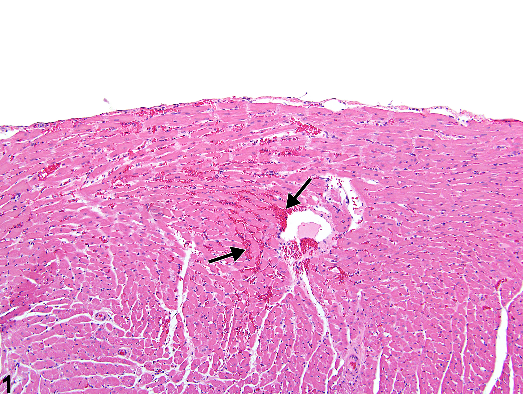
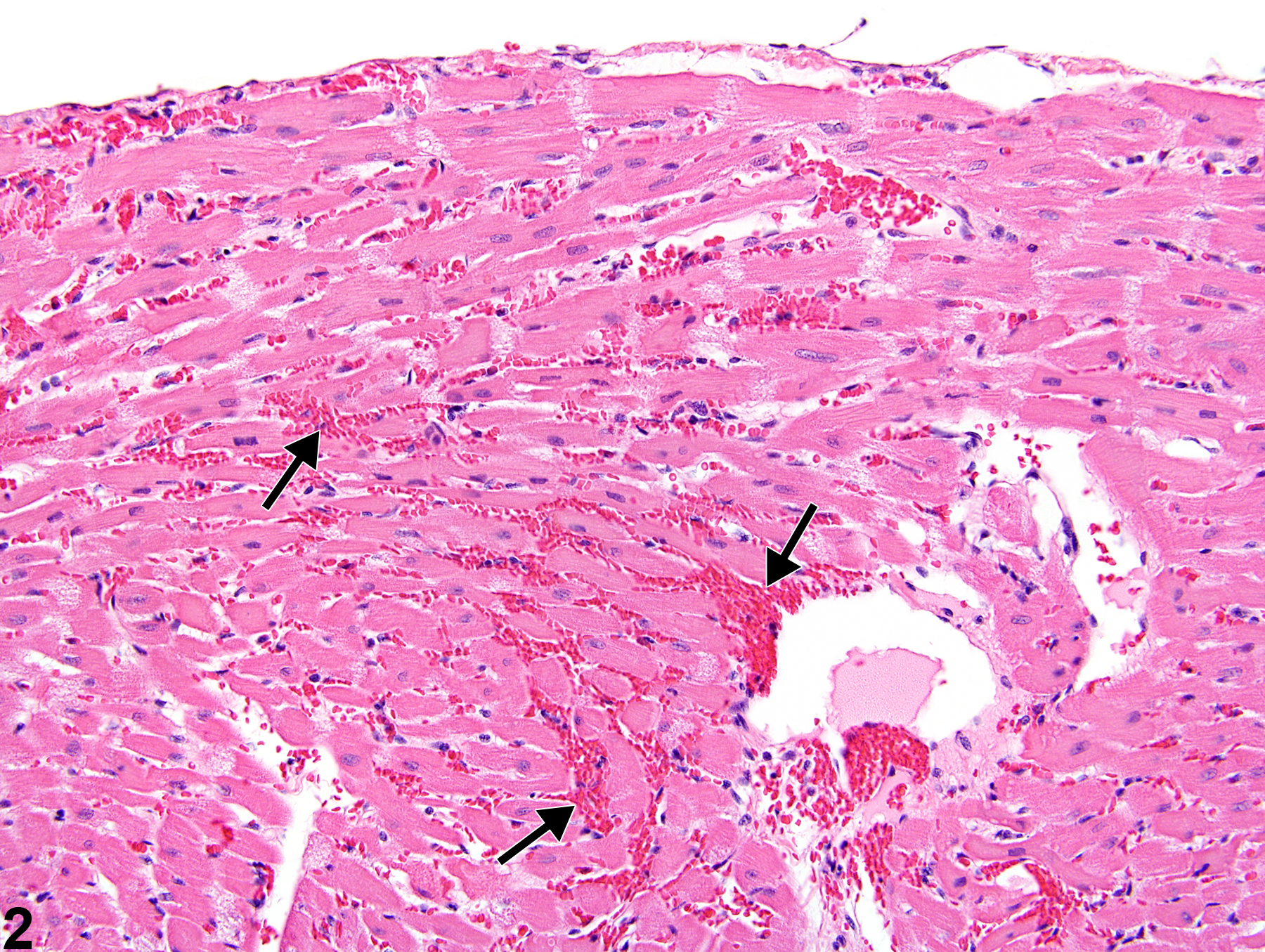
Heart - Hemorrhage in a male F344/N rat from an acute study. Extravasated erythrocytes are present in the myocardium (arrows).
Figure 1 of 4
Image of hemorrhage in the heart from a male F344/N rat in a acute study
Heart - Hemorrhage in a male F344/N rat from an acute study (higher magnification of Figure 1). Extravasated erythrocytes separate cardiomyocytes (arrows).
Figure 2 of 4
Image of hemorrhage in the heart from a male B6C3F1/N mouse in a subchronic study
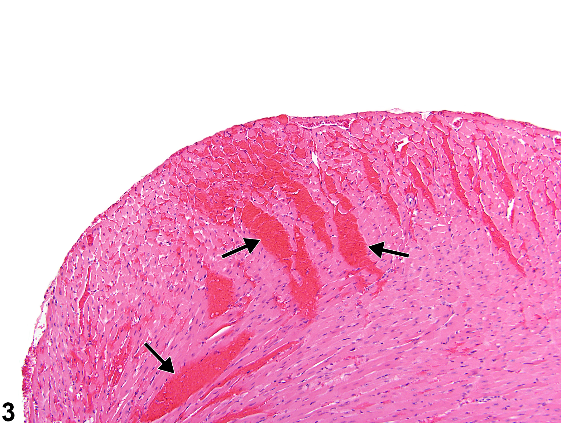
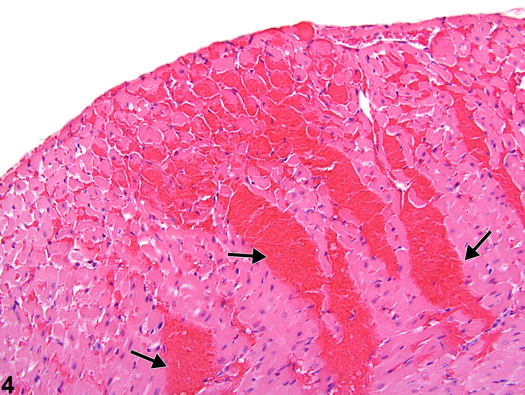
Heart - Hemorrhage in a male B6C3F1/N mouse from a subchronic study. Large areas of extravascular red blood cells are present in the myocardium (arrows).
Figure 3 of 4
Image of hemorrhage in the heart from a male B6C3F1/N mouse in a subchronic study
Heart - Hemorrhage in a male B6C3F1/N mouse from a subchronic study (higher magnification of Figure 3). Large areas of extravascular erythrocytes separate cardiomyocytes (arrows).
Figure 4 of 4
next arrow

comment:

Hemorrhage consists of few to many small, irregular foci of extravasated erythrocytes scattered within the myocardium ( Figure 1image opens in a pop-up window , Figure 2image opens in a pop-up window , Figure 3image opens in a pop-up window , and Figure 4image opens in a pop-up window ). Myocardial hemorrhage may be accompanied by other morphologic manifestations of myocardial damage (i.e., necrosis, macrophage infiltration). The presence of hemorrhage and cellular necrosis may indicate vascular damage or vasoconstriction of small arterial vessels, or may represent myocytic toxicity. Hemosiderin-laden macrophages may be seen.

recommendation:

Heart - Hemorrhage should be diagnosed and graded based on the extent of the lesion. The distribution of the hemorrhage (e.g., focal, multifocal, or diffuse), as well as the location (i.e., right ventricle, left ventricle, atrium, myocardium, papillary muscle), should be described in the narrative. If hemorrhage is secondary to another process such as inflammation or necrosis, it should not be diagnosed separately unless warranted by severity.

references:

Greaves P. 2000. Cardiovascular system. In: Histopathology of Preclinical Toxicity Studies (Greaves P, ed). Elsevier, Amsterdam, 254-311.

Jokinen MP, Boyle M, Lieuallen WG, Johnson CL, Malarkey DE, Nyska A. 2011. Morphologic aspects of rodent cardiotoxicity in a retrospective evaluation of National Toxicology Program studies. Toxicol Pathol 39(5):850-860.
Abstract: https://www.ncbi.nlm.nih.gov/pubmed/21747121

Nyska A, Murphy E, Foley JF, Collins BJ, Petranka J, Howden R, Hanlon P, Dunnick JK. 2005. Acute hemorrhagic myocardial necrosis and sudden death of rats exposed to a combination of ephedrine and caffeine. Toxicol Sci 83:388-396.
Abstract: https://www.ncbi.nlm.nih.gov/pubmed/15537744