

Cardiovascular System
Heart
Narrative
Examination of the heart is routinely included in toxicologic evaluation. In NTP studies, the heart is sectioned through the longitudinal axis, to include the right and left ventricles, interventricular septum, both atria, and a portion of the major vessels at the base of the heart.

Figure 1. Normal heart in a male B6C3F1/N mouse from a chronic study. Shown are the left ventricle (LV), right ventricle (RV), right and left atria (A), interventricular septum (IS), and aorta (asterisk). The pericardium is removed (not present).
The muscular chambers are composed of three layers: the epicardium, the myocardium, and the endocardium.
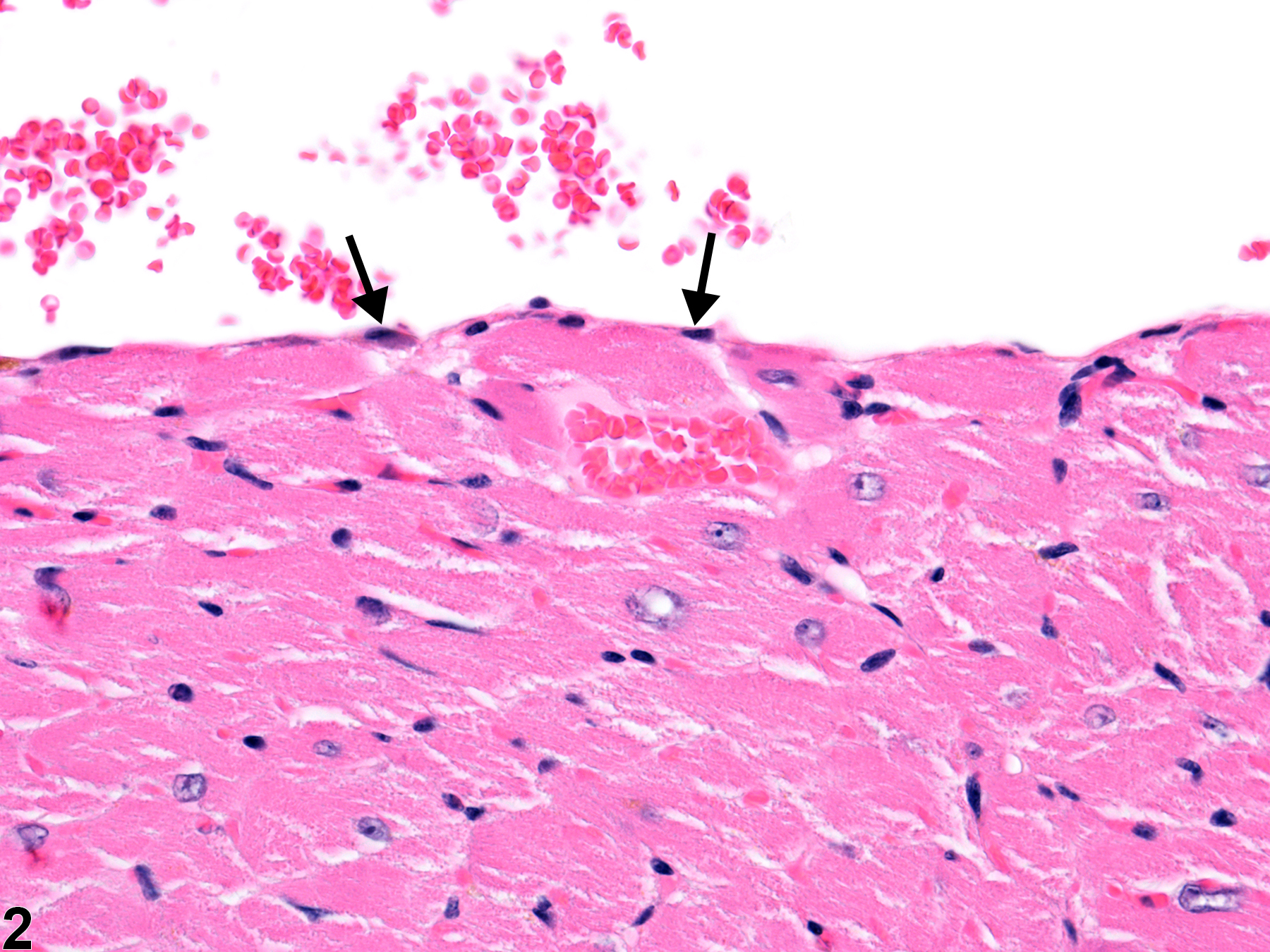
Figure 2. Normal heart in a male B6C3F1/N mouse from a chronic study. The outer endothelial layer is indicated by arrows.
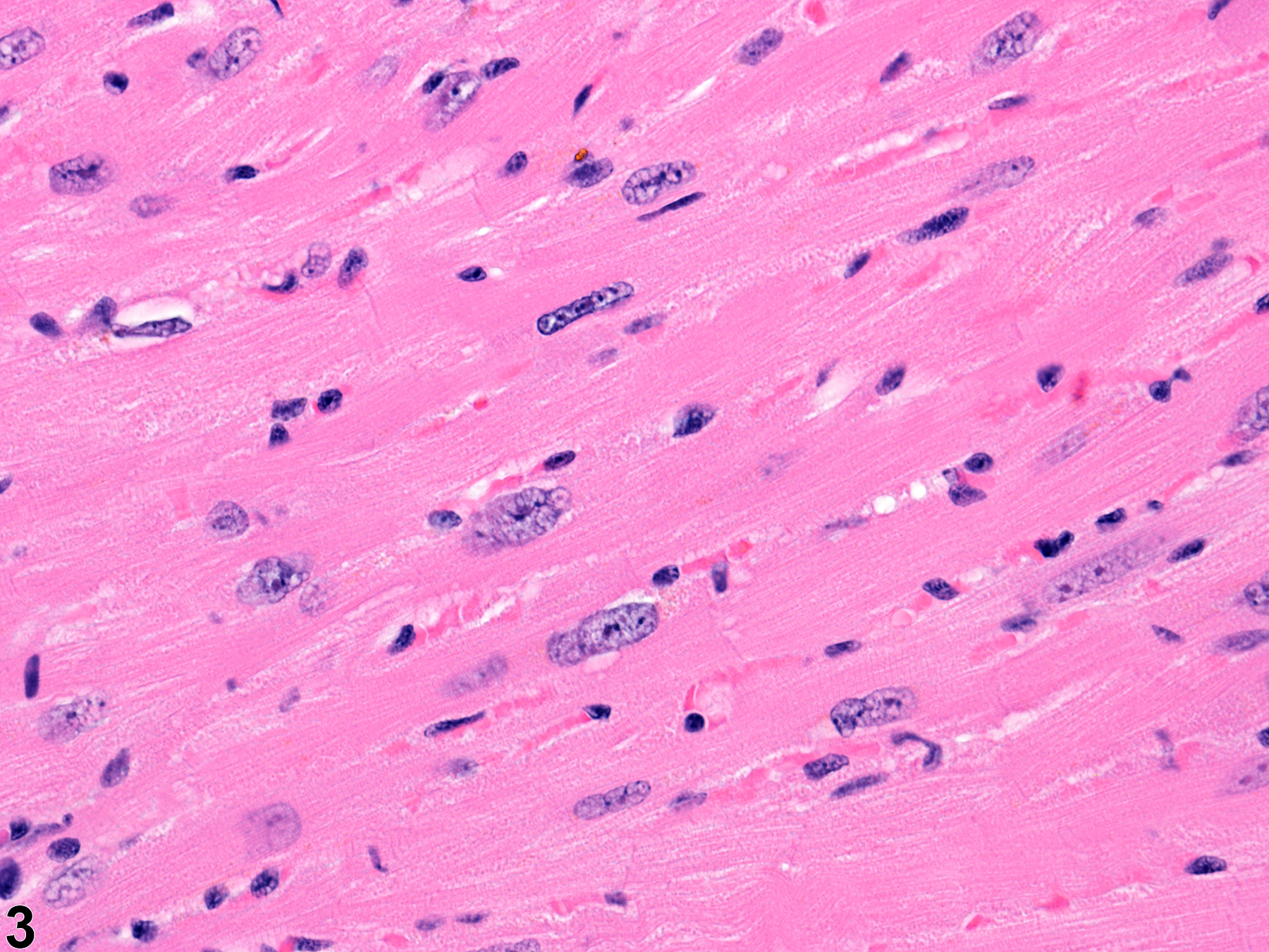
Figure 3. Normal myocardium in a male B6C3F1/N mouse from a chronic study.
Damage to the heart may be either structural or functional. Structural abnormalities may be detected microscopically, whereas functional changes are typically assessed through other means, such as enzyme detection or blood pressure measurements. Differentiating spontaneously occurring lesions from treatment-related lesions may be difficult. Spontaneous cardiomyopathy is commonly found in rats but less commonly in mice. Further, some spontaneous lesions may be exacerbated by treatment, making differentiation even more difficult.
Jokinen MP, Boyle M, Lieuallen WG, Johnson CL, Malarkey DE, Nyska A. 2011. Morphologic aspects of rodent cardiotoxicity in a retrospective evaluation of National Toxicology Program studies. Toxicol Pathol 39(5):850-860.
Abstract: https://www.ncbi.nlm.nih.gov/pubmed/21747121McKenzie WF, Alison R. 1990. Heart. In: Pathology of the Fischer Rat: Reference and Atlas (Boorman GA, Eustis SL, Elwell MR, Montgomery CA, MacKenzie WF, eds). Academic Press, San Diego, CA, 461-472.
Elwell MR, Mahler JF. 1999. Heart, blood vessels, and lymphatic vessels. In: Pathology of the Mouse: Reference and Atlas (Maronpot RR, Boorman GA, Gaul BW, eds). Cache River Press, Vienna, IL, 361-380.
Wallig MA, Haschek WM, Rousseaux CG, eds. 2013. Cardiac, vascular, and skeletal muscle systems. In: Handbook of Toxicologic Pathology, 3rd ed. Academic Press, San Diego, CA, 1567-1586.