U.S. flag

An official website of the United States government

Dot gov

The .gov means it's official.
Federal government websites often end in .gov or .mil. Before sharing sensitive information, make sure you're on a federal government site.

Https

The site is secure.
The https:// ensures that you are connecting to the official website and that any information you provide is encrypted and transmitted securely.

Skip to Main Navigation
Skip to Page Content
Skip to Atlas Navigation

Heart - Mineral

Image of mineral in the heart from a male F344/N rat in a chronic study
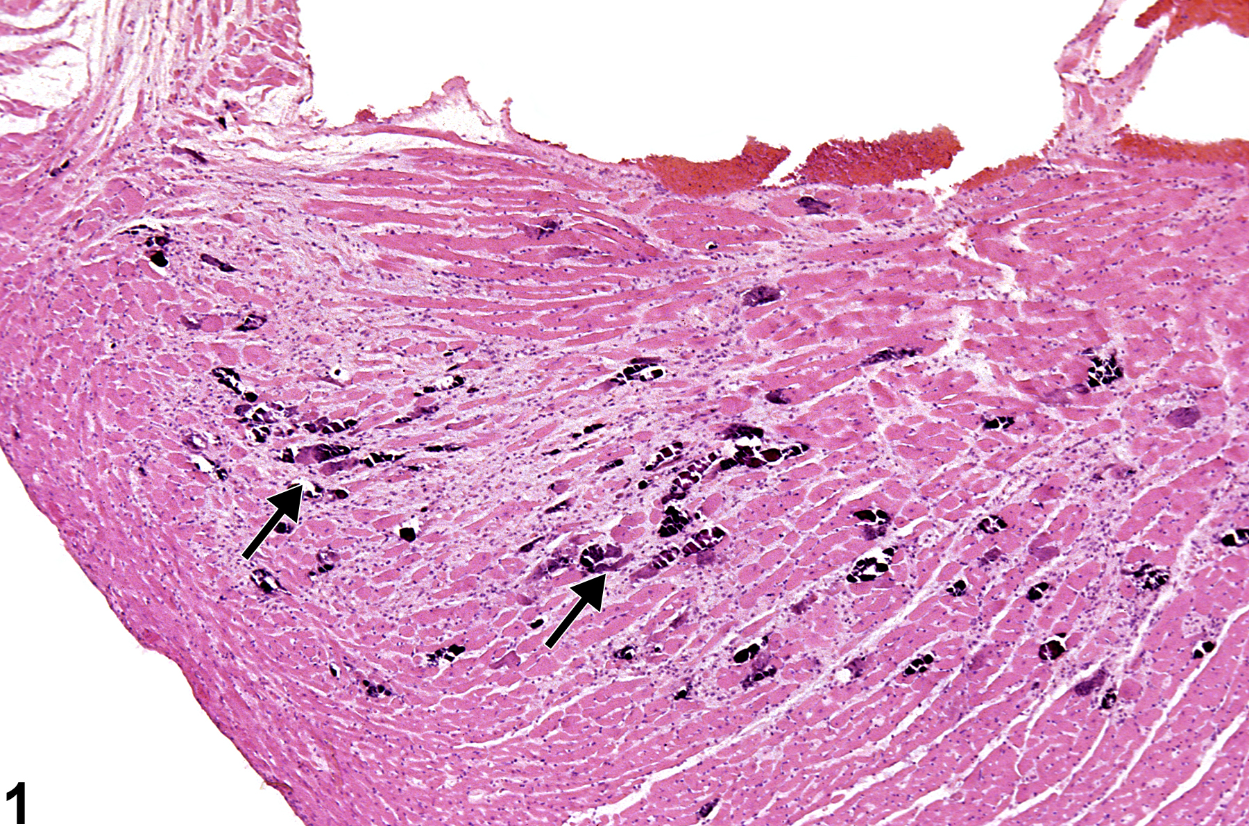
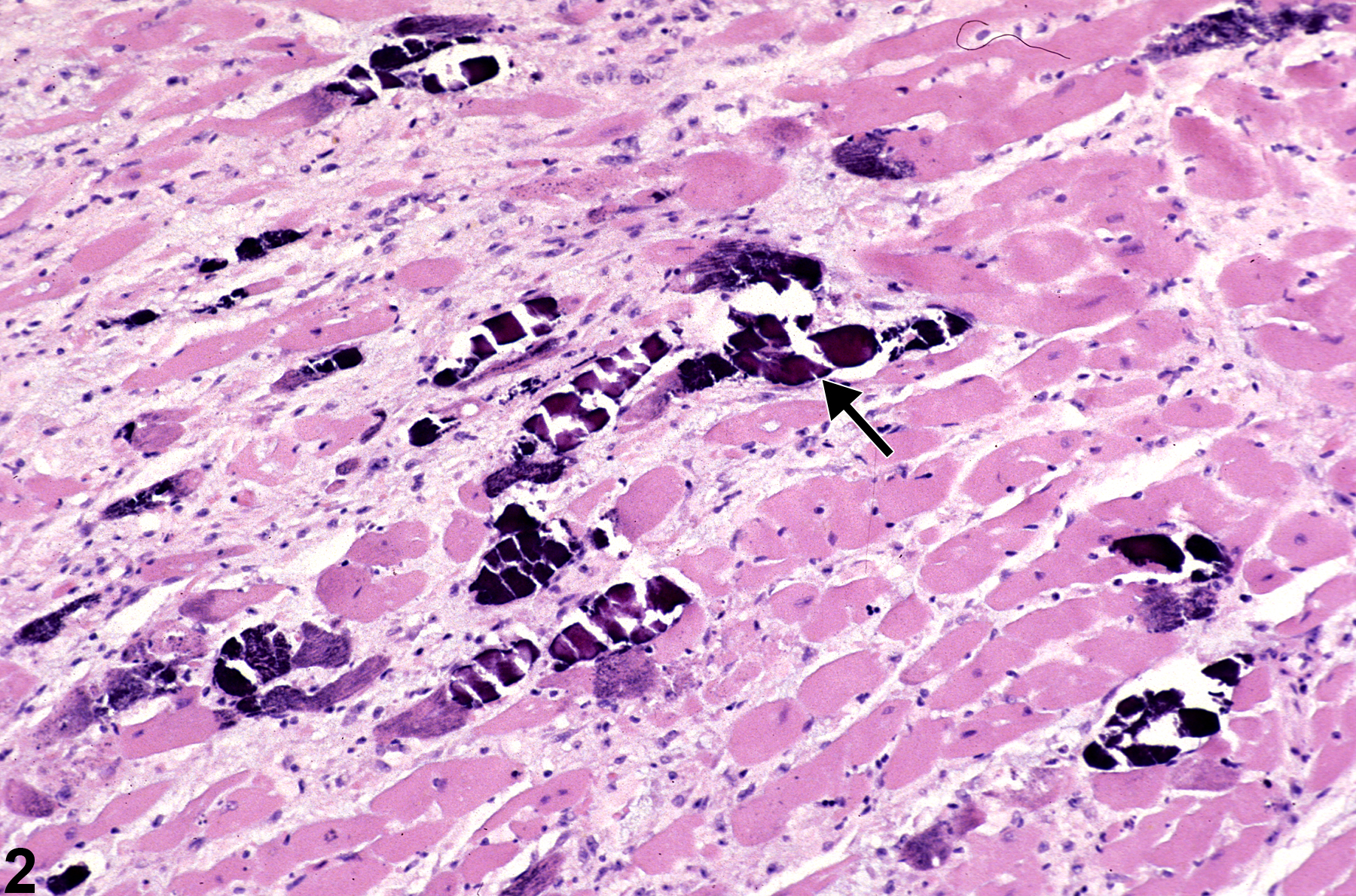
Heart - Mineral in a male F344/N rat from a chronic study. Dark blue crystalline material is present in the myocardium (arrows).
Figure 1 of 4
Image of mineral in the heart from a male F344/N rat in a chronic study
Heart - Mineral in a male F344/N rat from a chronic study (higher magnification of Figure 1). The dark blue mineral (arrow) in the cardiomyocytes is associated with a region of fibrosis.
Figure 2 of 4
Image of mineral in the heart from a female B6C3F1/N mouse in a subchronic study
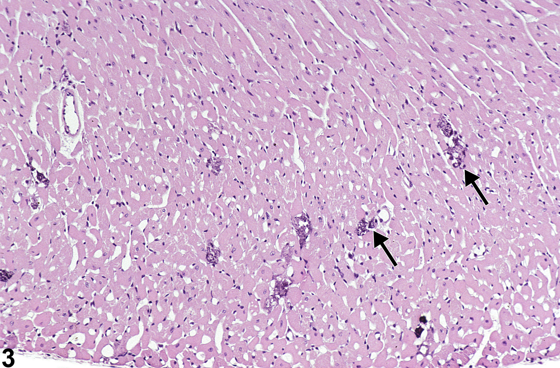
Heart - Mineral in a female B6C3F1/N mouse from a subchronic study. Focal areas of mineral are present in the myocardium (arrows).
Figure 3 of 4
Image of mineral in the heart from a female B6C3F1/N mouse in a subchronic study
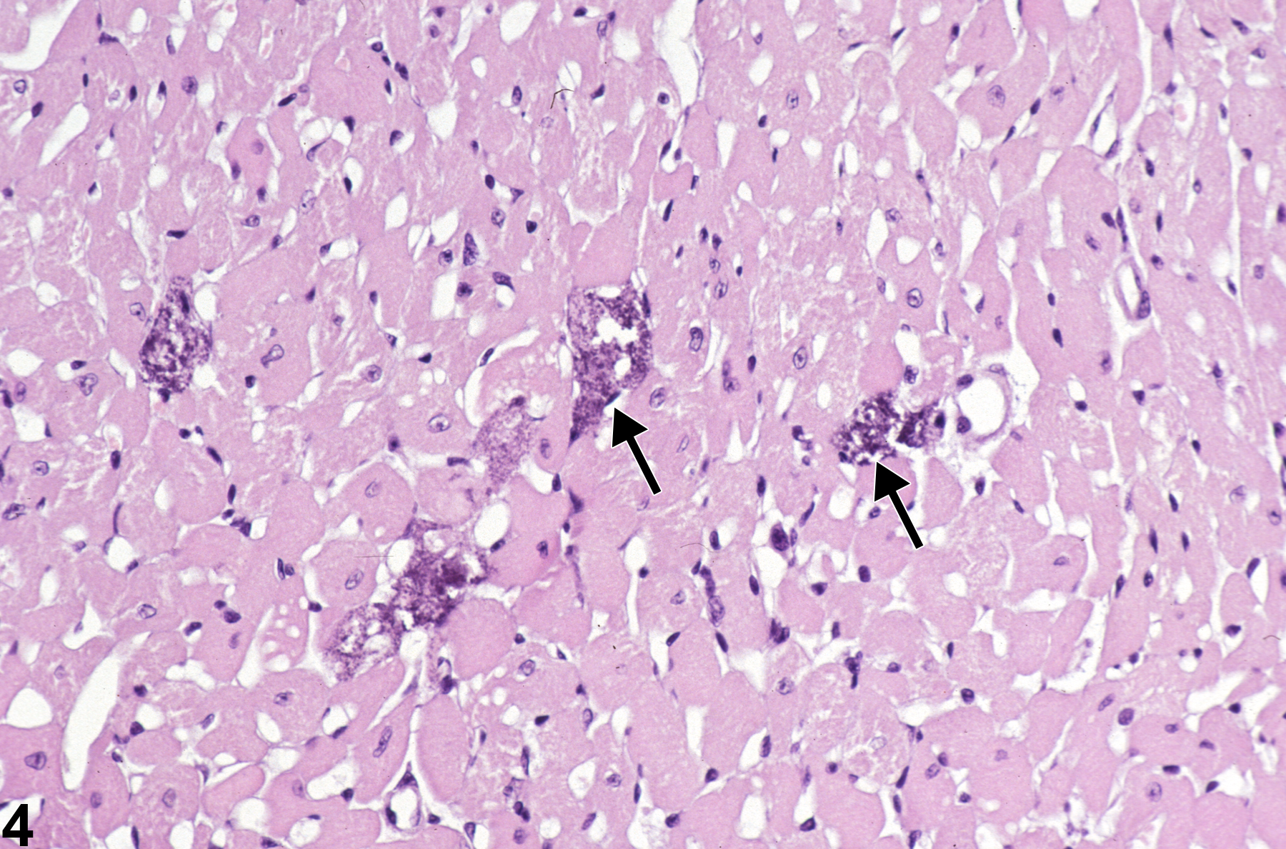
Heart - Mineral in a female B6C3F1/N mouse from a subchronic study (higher magnification of Figure 3). There is blue mineral within the cardiomyocytes.
Figure 4 of 4
next arrow

comment:

Microscopically, mineral ( Figure 1image opens in a pop-up window , Figure 2image opens in a pop-up window , Figure 3image opens in a pop-up window , and Figure 4image opens in a pop-up window ) ranges from light basophilic stippling in slightly affected myofibers to intense, diffuse basophilic granularity that completely obscures the sarcoplasm in severely affected cardiomyocytes. The mineralization may be localized within individual cardiomyocytes, associated with sites of cellular necrosis (dystrophic), or found within the interstitial connective tissue or epicardium. Cardiac mineralization may be a spontaneous change (e.g., genetically or age related), a manifestation of primary cardiotoxicity (dystrophic mineralization), or secondary to renal disease (metastatic mineralization).

BALB/c mice develop epicardial mineralization, predominantly of the right ventricle. C3H mice develop myocardial mineralization of both ventricles. DBA mice may develop epicardial and myocardial mineralization.

recommendation:

Whenever present, mineral in the heart should be recorded and graded based on the extent of the mineralized deposits. Special stains are generally not needed for confirmation of mineral deposition. The location of the lesion (i.e., myocardium, epicardium) should be described in the narrative. If possible, differentiate dystrophic from metastatic mineral and discuss if changes are present secondary to renal disease (metastatic mineralization). Because it is secondary to necrosis, dystrophic mineralization should not be diagnosed separately, unless its severity warrants a separate diagnosis. However, its presence may be noted in the pathology narrative.

references:

Jokinen MP, Boyle M, Lieuallen WG, Johnson CL, Malarkey DE, Nyska A. 2011. Morphologic aspects of rodent cardiotoxicity in a retrospective evaluation of National Toxicology Program studies. Toxicol Pathol 39(5):850-860.
Abstract: https://www.ncbi.nlm.nih.gov/pubmed/21747121