

Endocrine System
Adrenal Gland - Angiectasis
Narrative
Angiectasis can be associated with or secondary to other adrenal lesions such as inflammation, atrophy, degeneration, and neoplasia. It can also occur as a spontaneous age-related change in old rats and mice.
Angiectasis and hemangioma can be difficult to differentiate, but a distinction should be attempted. Hemangiomas tend to be well-circumscribed, unencapsulated masses composed of densely packed, dilated vascular spaces. The vascular spaces are lined by a single layer of normal-appearing endothelial cells aligned on usually delicate collagenous septa, though some cases exhibit more abundant collagenous stroma. In contrast, although angiectasis is also characterized by dilated vascular channels lined by unremarkable endothelium, the vascular channels are more loosely or irregularly arranged rather than presenting as a well-demarcated mass.
Hamlin MH, Banas DA. 1990. Adrenal gland. In: Pathology of the Fischer Rat: Reference and Atlas (Boorman GA, Eustis SL, Elwell MR, Montgomery CA, MacKenzie WF, eds). Academic Press, San Diego, 501-518.
Abstract: https://www.ncbi.nlm.nih.gov/nlmcatalog/9002563McInnes EF. 2011. Wistar and Sprague Dawley rats. In: Background Lesions in Laboratory Animals: A Color Atlas (McInnes EF, ed). Saunders Elsevier, Amsterdam, 16-36.
Abstract: http://www.sciencedirect.com/science/book/9780702035197National Toxicology Program. 1996. NTP TR-455. Toxicology and Carcinogenesis Studies of Codeine (CAS No. 76-57-3) in F344 Rats and B6C3F1 Mice (Feed Studies). NTP, Research Triangle Park, NC.
Abstract: https://ntp.niehs.nih.gov/go/6054Rosol TJ, Yarrington JT, Latendresse J, Capen CC. 2001. Adrenal gland: Structure, function, and mechanisms of toxicity. Toxicol Pathol 29:41-48.
Abstract: https://www.ncbi.nlm.nih.gov/pubmed/11215683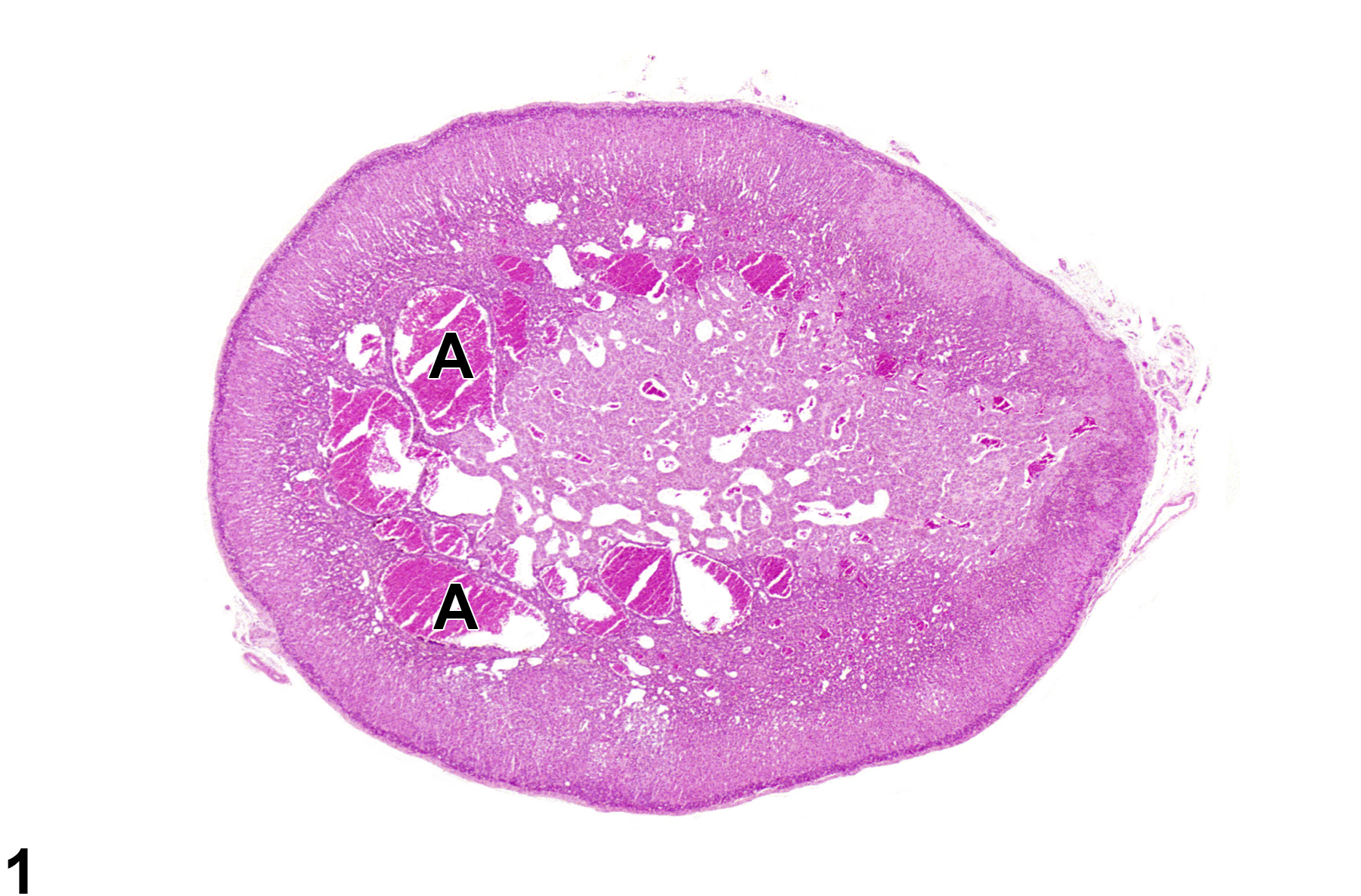
Adrenal gland, Cortex - Angiectasis in a male F344/N rat from a chronic study. There are widely dilated, blood-filled vascular channels (A) in the cortex of the adrenal gland.
All Images

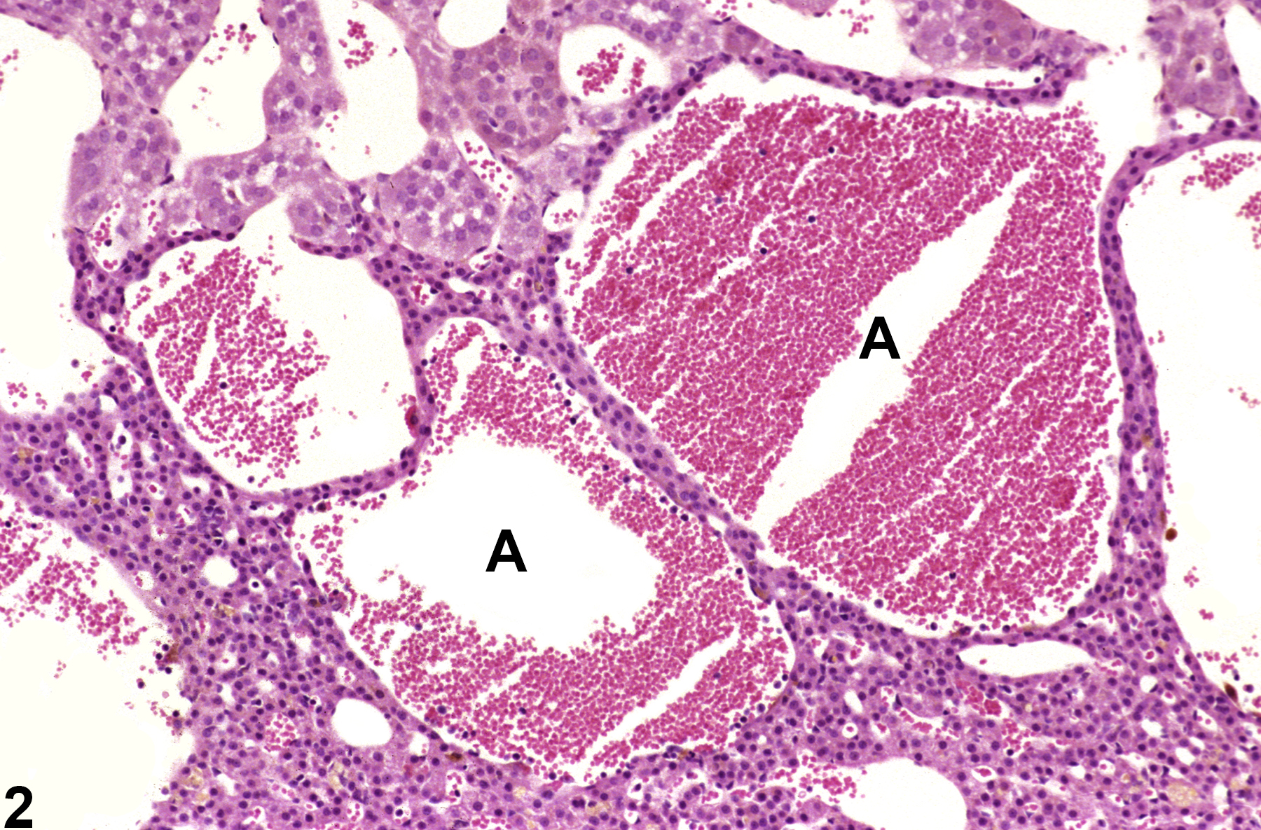
Adrenal gland, Cortex - Angiectasis in a male F344/N rat from a chronic study. There are widely dilated, blood-filled vascular channels (A) in the cortex of the adrenal gland.

Adrenal gland, Cortex - Angiectasis in a male F344/N rat from a chronic study (higher magnification of Figure 1). The widely dilated vascular channels (A) cause slight compression of adjacent tissue.