U.S. flag

An official website of the United States government

Dot gov

The .gov means it's official.
Federal government websites often end in .gov or .mil. Before sharing sensitive information, make sure you're on a federal government site.

Https

The site is secure.
The https:// ensures that you are connecting to the official website and that any information you provide is encrypted and transmitted securely.

Skip to Main Navigation
Skip to Page Content
Skip to Atlas Navigation

Adrenal Gland - Cyst

Image of cyst in the adrenal gland from a female B6C3F1/N mouse in a chronic study
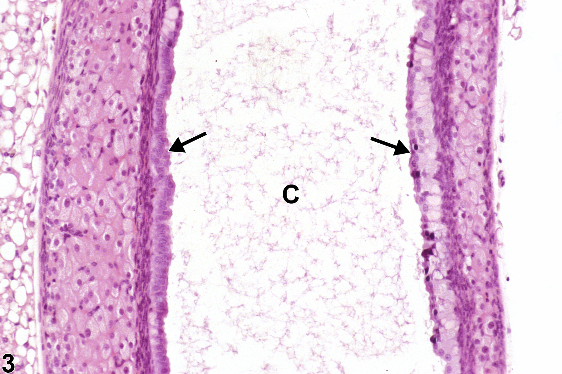
Adrenal gland, Cortex - Cyst in a female B6C3F1/N mouse from a chronic study. An adrenal cortical cyst (C), filled with amorphous pale eosinophilic material, compresses adjacent cortex and medulla.
Figure 1 of 3
Image of cyst in the adrenal gland from a female B6C3F1/N mouse in a chronic study
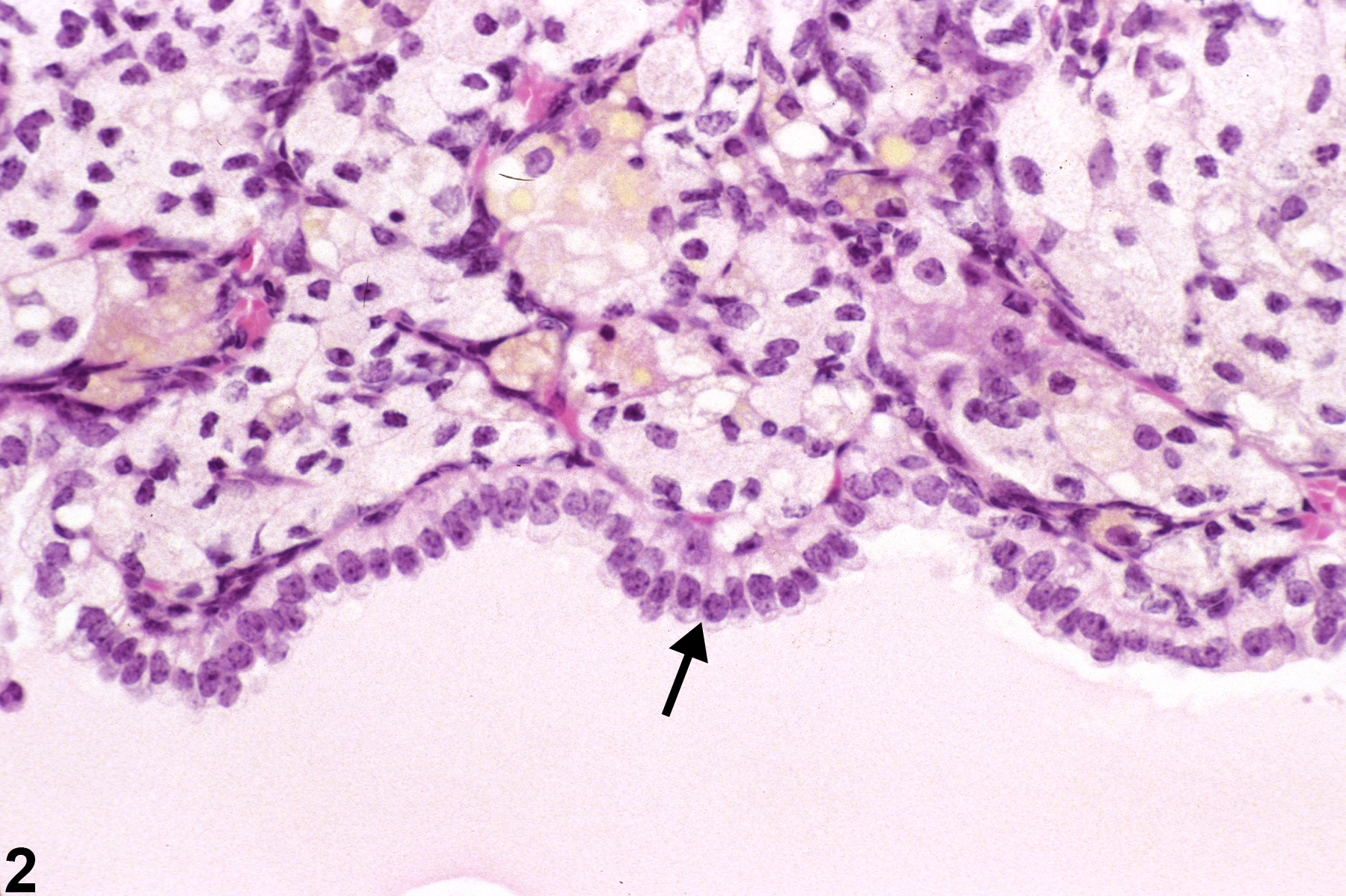
Adrenal gland, Cortex - Cyst in a female B6C3F1/N mouse from a chronic study (higher magnification of Figure 1). A single layer of well-differentiated, low columnar epithelial cells (arrow) lines an adrenocortical cyst.
Figure 2 of 3
Image of cyst in the adrenal gland from a female B6C3F1/N mouse in a chronic study
Adrenal gland, Cortex - Cyst in a female B6C3F1/N mouse from a chronic study. Cortical cyst (C) lined by cuboidal to columnar epithelial cells (arrows) contains flocculent, pale eosinophilic material.
Figure 3 of 3
next arrow

comment:

Adrenal cysts ( Figure 1image opens in a pop-up window , Figure 2image opens in a pop-up window , and Figure 3image opens in a pop-up window ) are lined by cuboidal to ciliated columnar epithelium and may contain amorphous to flocculent, eosinophilic material. They may be located in the cortex or medulla and may cause variable distortion or compression of the adjacent tissue. These minor findings are more common in mice than in rats and are usually incidental, spontaneous changes of no clinical or toxicologic relevance. Cortical cysts must be distinguished from angiectasis (dilated vascular channels containing blood and lined by flat endothelial cells). Cortical cysts in rats must also be distinguished from cystic degeneration, a common lesion in certain rat strains, which features irregular, often large cavitated spaces filled with proteinaceous fluid but lack the epithelial lining of true cysts ( Figure 2image opens in a pop-up window and Figure 3image opens in a pop-up window ).

recommendation:

Adrenal gland cysts should be diagnosed only if they occur with treatment-related increased incidences. In these cases, they should be diagnosed but not assigned of a severity grade. When diagnosed, an appropriate site modifier (i.e., cortex or medulla) should be included in the diagnosis to indicate the location of the cyst.

references:

National Toxicology Program. 1993. NTP TR-434. Toxicology and Carcinogenesis Studies of 1,3-Butadiene (CAS No. 106-99-0) in B6C3F1 Mice (Inhalation Studies). NTP, Research Triangle Park, NC.
Abstract: https://ntp.niehs.nih.gov/go/6012

Nyska A, Maronpot RR. 1990. Adrenal gland. In: Pathology of the Mouse: Reference and Atlas (Maronpot RR, Boorman GA, Gaul BW, eds). Cache River Press, Vienna, IL, 509-536.
Abstract: http://www.cacheriverpress.com/books/pathmouse.htm