

Endocrine System
Adrenal Gland - Extramedullary Hematopoiesis
Narrative
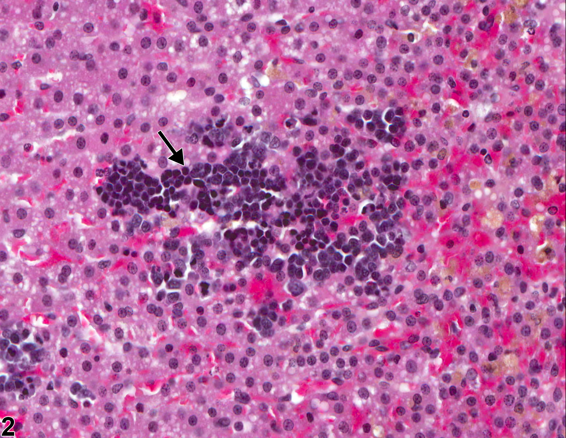
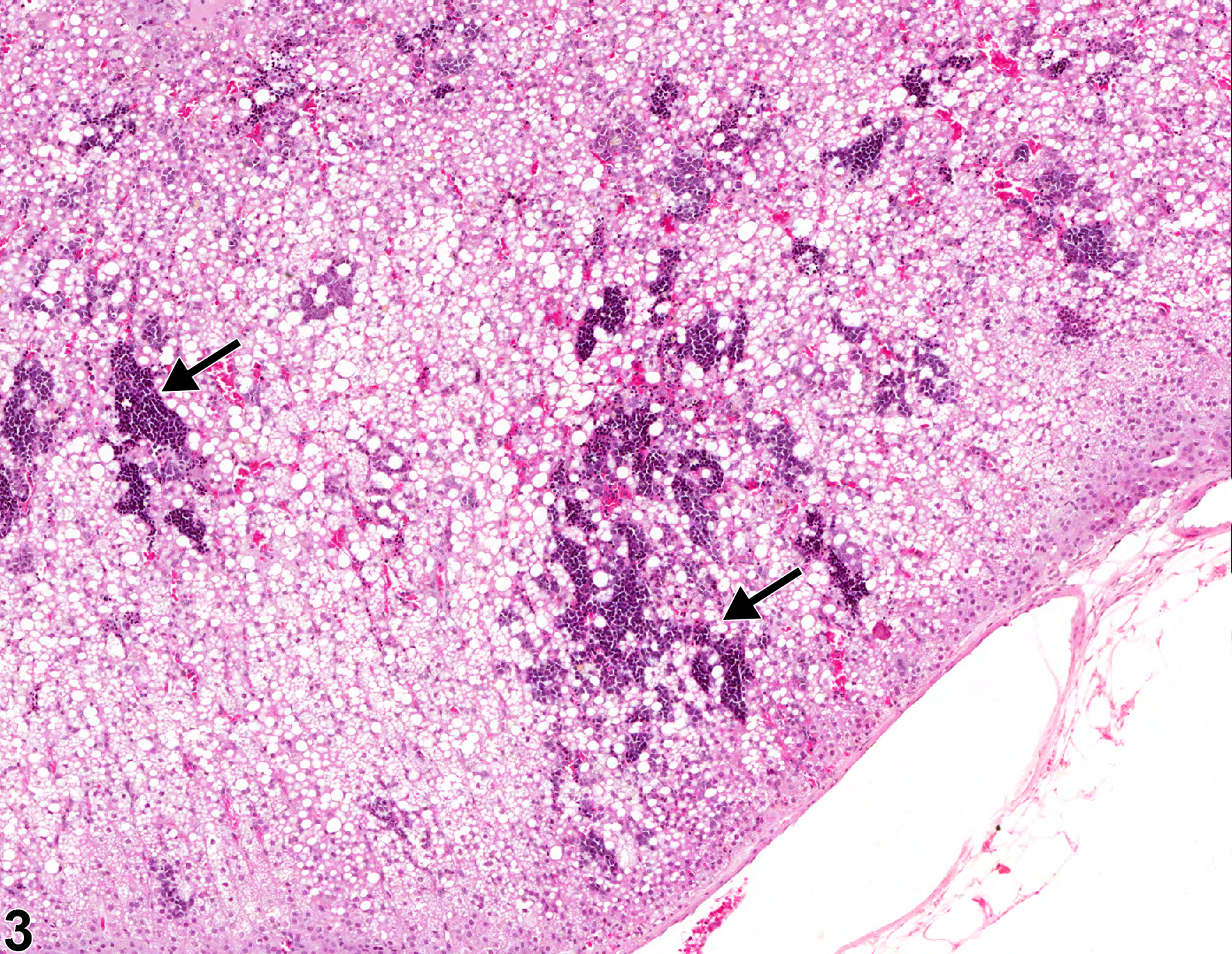
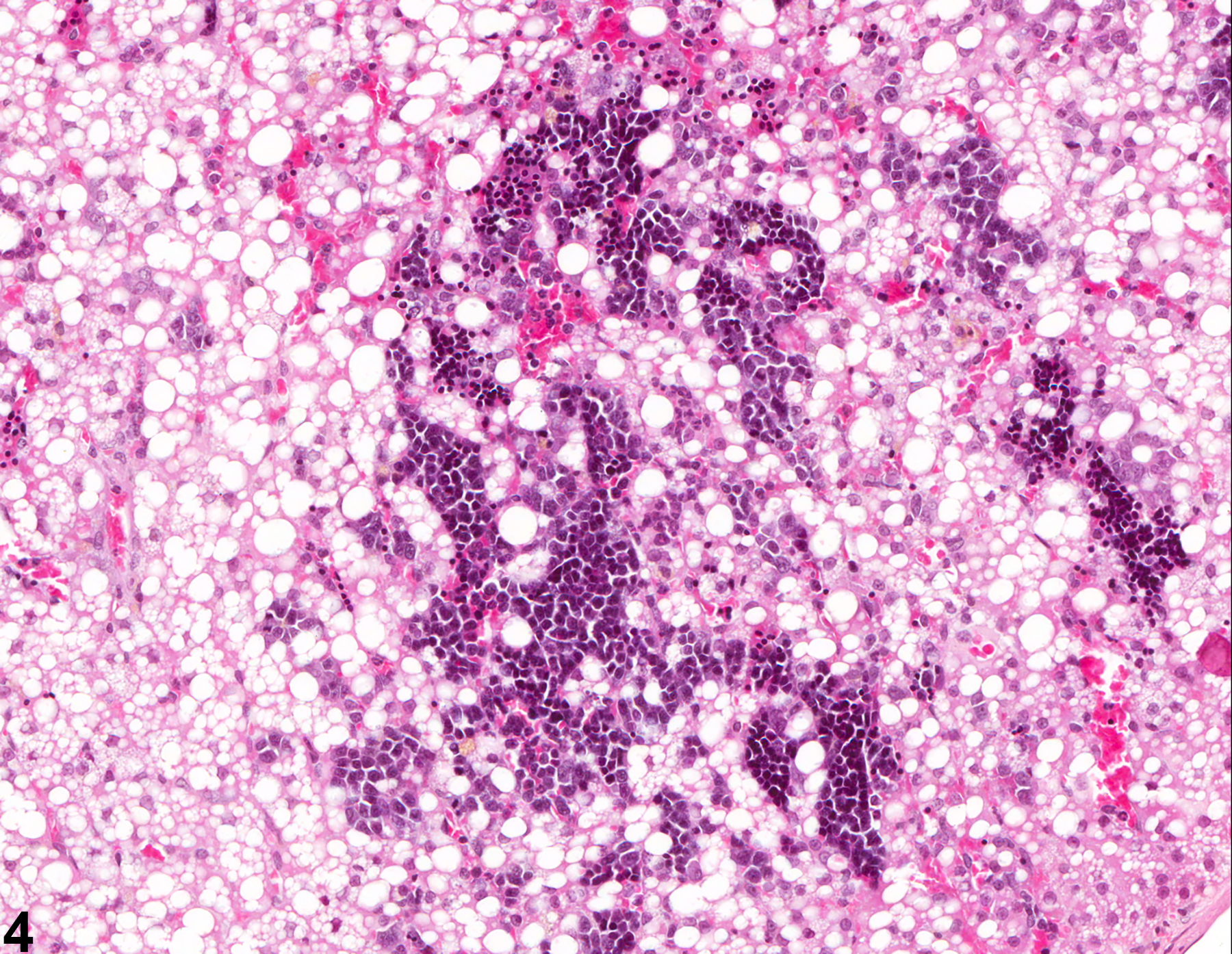
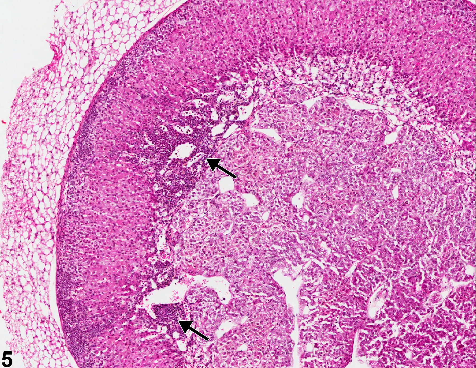
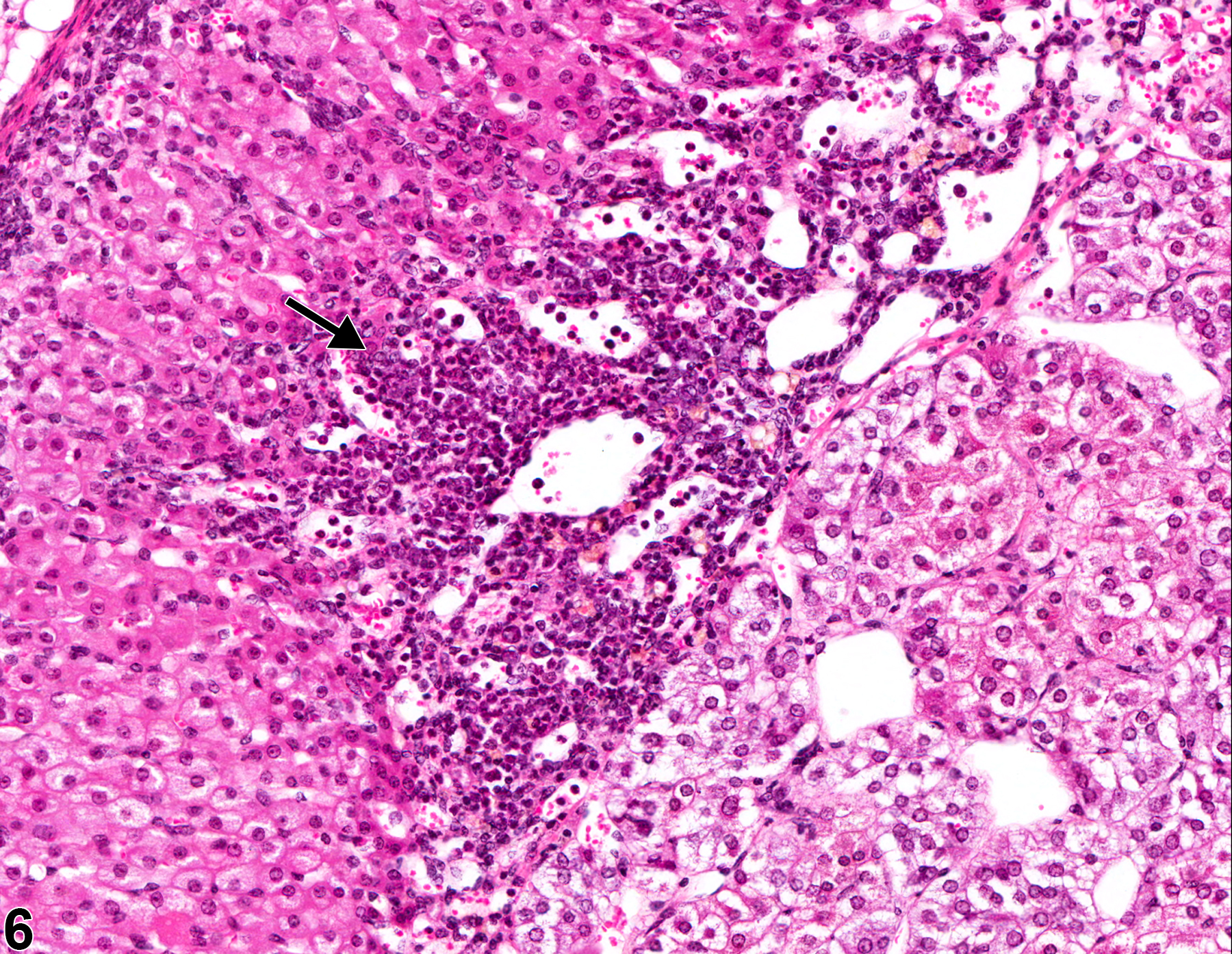
Adrenal EMH consists of variably sized, noncompressive clusters of immature hematopoietic cells and/or megakaryocytes scattered throughout the cortex and/or medulla (Figure 1, Figure 2, Figure 3, Figure 4, Figure 5, and Figure 6). Myelopoietic cells are typically larger than erythropoietic cells and have more cytoplasm, whereas erythropoietic cells typically are much darker. Adrenal EMH must be distinguished from inflammation (mature leukocytes, often associated with tissue necrosis or degeneration) and from systemic lymphoid or hematopoietic neoplastic infiltrates (poorly differentiated, often anaplastic cells usually in extensive accumulations that distort or obliterate normal architecture).
Frith CH, Botts S, Jokinen MP, Eighmy JJ, Hailey JR, Morgan SJ, Chandra M. 2000. Non-proliferative lesions of the endocrine system in rats, E-1. In: Guides for Toxicologic Pathology. STP/ARP/AFIP, Washington, DC.
Full Text: https://www.toxpath.org/docs/SSNDC/EndocrineNonprolifRat.pdfHamlin MH, Banas DA. 1990. Adrenal gland. In: Pathology of the Fischer Rat: Reference and Atlas (Boorman GA, Eustis SL, Elwell MR, Montgomery CA, MacKenzie WF, eds). Academic Press, San Diego, 501-518.
Abstract: https://www.ncbi.nlm.nih.gov/nlmcatalog/9002563Johns JL, Christopher MM. 2012. Extramedullary hematopoiesis: A new look at underlying stem cell niche, theories of development, and occurrence in animals. Vet Pathol 49:508-523.
Abstract: https://www.ncbi.nlm.nih.gov/pubmed/22262354McInnes EF. 2011. Wistar and Sprague Dawley rats. In: Background Lesions in Laboratory Animals: A Color Atlas (McInnes EF, ed). Saunders Elsevier, Amsterdam, 16-36.
Abstract: http://www.sciencedirect.com/science/book/9780702035197National Toxicology Program. 1993. NTP TR-402. Toxicology and Carcinogenesis Studies of Furan (CAS No. 110-00-9) in F344 Rats and B6C3F1 Mice (Gavage Studies). NTP, Research Triangle Park, NC.
Abstract: https://ntp.niehs.nih.gov/go/12255National Toxicology Program. 1993. NTP TR-434. Toxicology and Carcinogenesis Studies of 1,3-Butadiene (CAS No. 106-99-0) in B6C3F1 Mice (Inhalation Studies). NTP, Research Triangle Park, NC.
Abstract: https://ntp.niehs.nih.gov/go/6012National Toxicology Program. 1997. NTP TR-463. Toxicology and Carcinogenesis Studies of D&C Yellow No. 11 (CAS No. 8003-22-3) in F344/N Rats (Feed Studies). NTP, Research Triangle Park, NC.
Abstract: https://ntp.niehs.nih.gov/go/6070Nyska A, Maronpot RR. 1990. Adrenal gland. In: Pathology of the Mouse: Reference and Atlas (Maronpot RR, Boorman GA, Gaul BW, eds). Cache River Press, Vienna, IL, 509-536.
Taylor I. 2011. Mouse. In: Background Lesions in Laboratory Animals: A Color Atlas (McInnes EF, ed). Saunders Elsevier, Amsterdam, 45-72.
Abstract: http://www.sciencedirect.com/science/book/9780702035197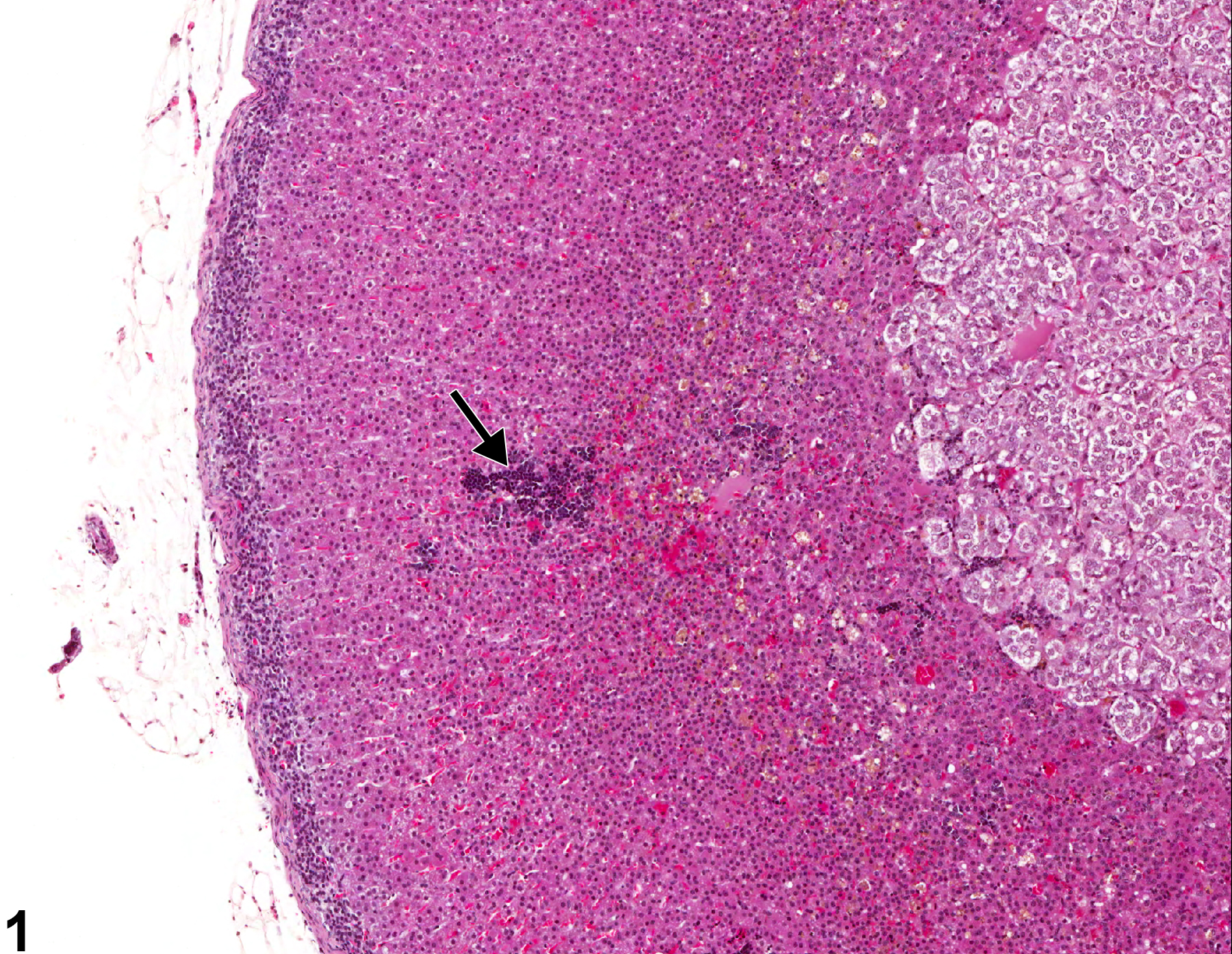
Adrenal gland, Cortex - Extramedullary hematopoiesis in a female F344/N rat from a chronic study. There is a focus of hematopoietic cells (arrow) in the cortex (zona fasciculata).
All Images

Adrenal gland, Cortex - Extramedullary hematopoiesis in a female F344/N rat from a chronic study. There is a focus of hematopoietic cells (arrow) in the cortex (zona fasciculata).

Adrenal gland, Cortex - Extramedullary hematopoiesis in a female F344/N rat from a chronic study (higher magnification of Figure 1). There is a focus of immature hematopoietic cells (arrow) in the sinusoids of the cortex (zona fasciculata).

Adrenal gland, Cortex - Extramedullary hematopoiesis in a male F344/N rat from a chronic study. There are multiple foci of hematopoietic cells (arrows) in the adrenal zona fasciculata.

Adrenal gland, Cortex - Extramedullary hematopoiesis in a male F344/N rat from a chronic study (higher magnification of Figure 3). There is a focus of immature hematopoietic cells (arrow) within the sinusoids of the zona fasciculata.

Adrenal gland, Cortex - Extramedullary hematopoiesis in a female B6C3F1/N mouse from a chronic study. There are several foci of hematopoietic cells (arrows) at the corticomedullary junction.

Adrenal gland, Cortex - Extramedullary hematopoiesis in a female B6C3F1/N mouse from a chronic study (higher magnification of Figure 5). A focus of immature hematopoietic cells (arrow) is present at the corticomedullary junction.