

Endocrine System
Adrenal Gland - Inflammation
Narrative
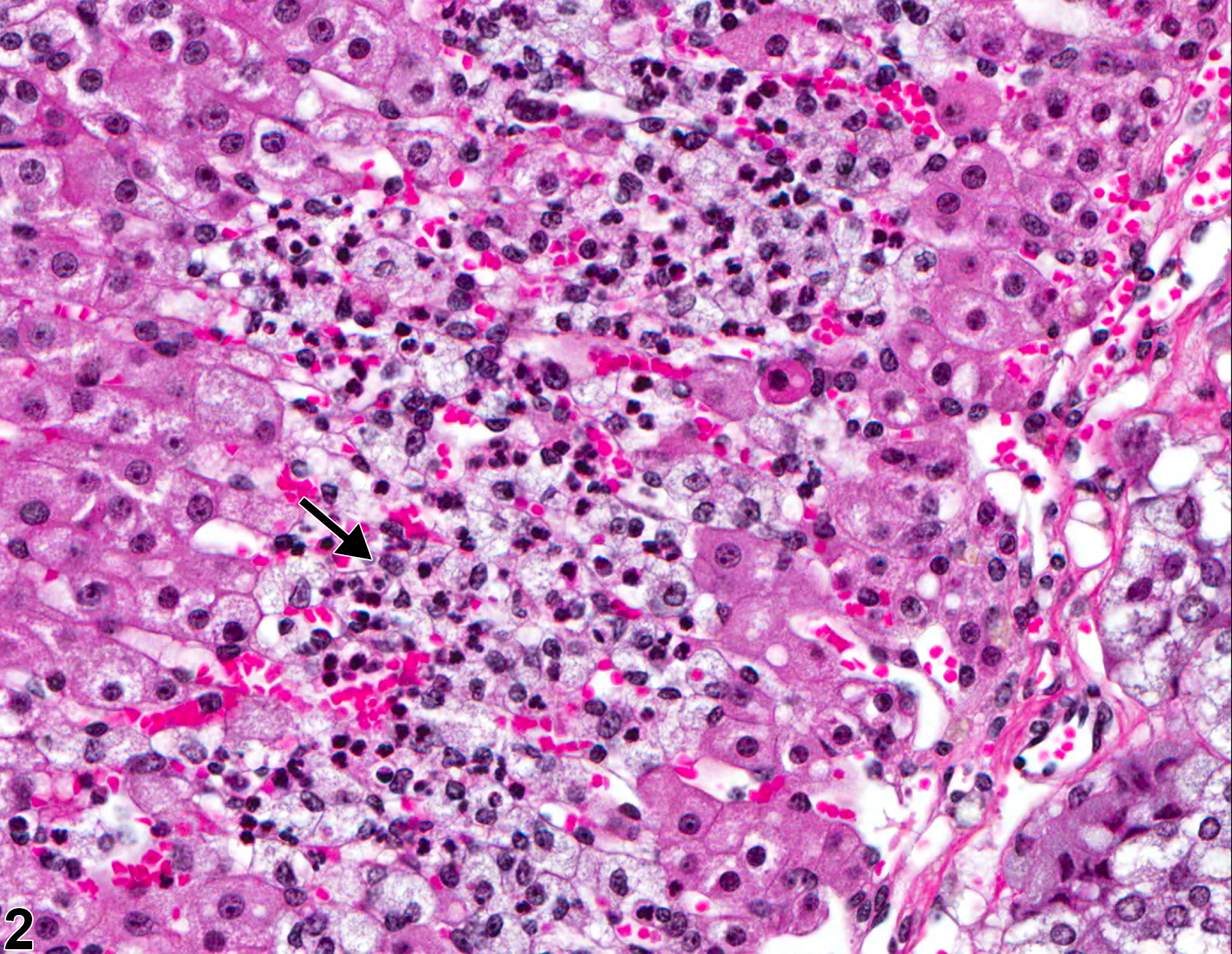
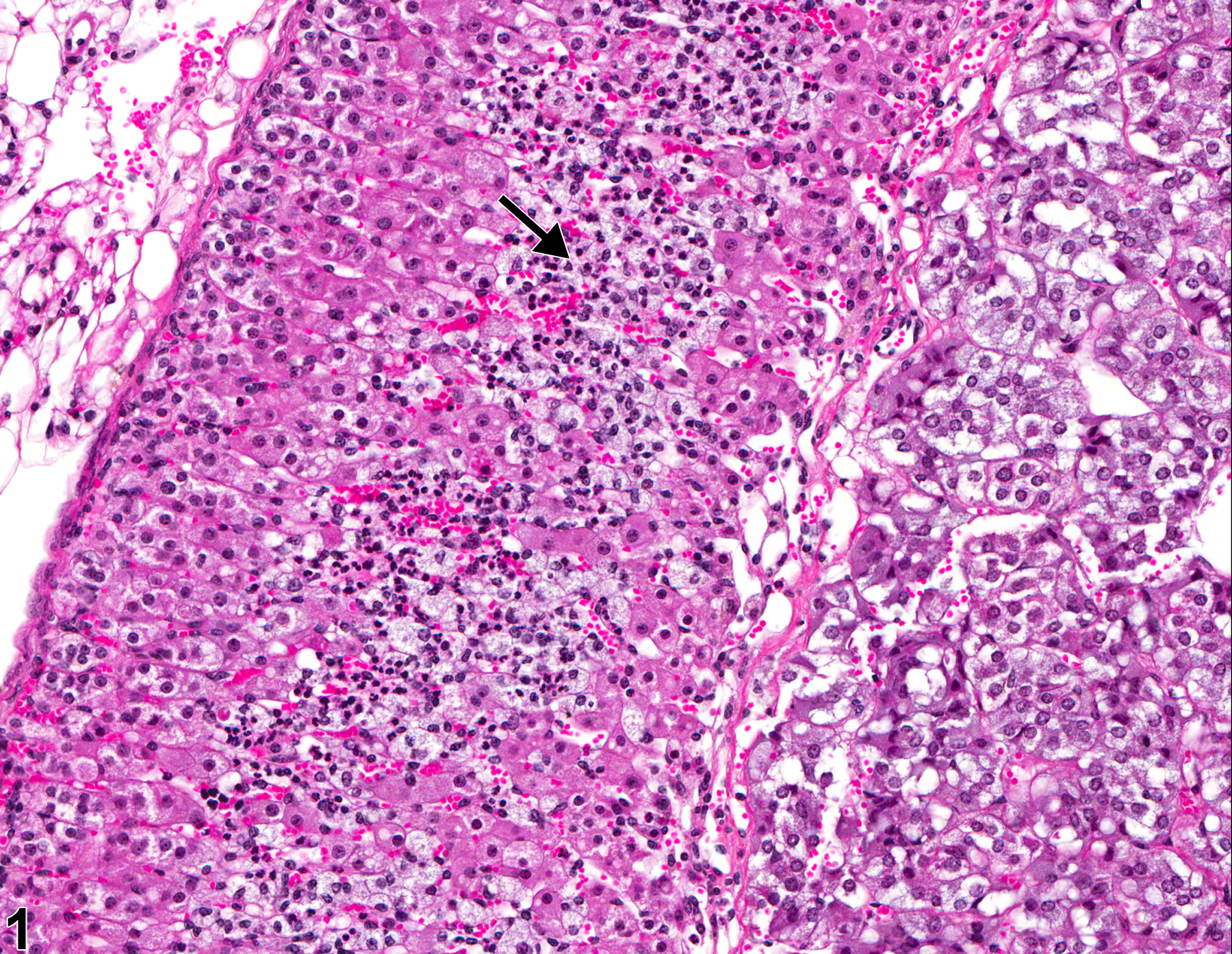
In NTP studies, there are five standard categories of inflammation: acute, suppurative, chronic, chronic-active, and granulomatous. Acute inflammation is characterized by infiltration of neutrophils (Figure 1 and Figure 2), which may be accompanied by eosinophils and macrophages, and occasional mast cells, lymphocytes, and plasma cells. Suppurative inflammation is characterized by discrete pockets of degenerate neutrophils and cellular debris. There may be evidence of chronicity, such as fibrosis and lymphoplasmacytic infiltrates, surrounding these pockets. Chronic inflammation is characterized by the presence of mononuclear cells (lymphocytes, macrophages, and plasma cells) and may be accompanied by fibrosis. Chronic-active inflammation is characterized by coexistence of elements of chronic inflammation (lymphocytes, macrophages, fibrosis) and superimposed acute inflammation (neutrophilic and/or eosinophilic infiltrates). Granulomatous inflammation is characterized by accumulations of macrophages, multinucleated giant cells, and variable numbers of lymphocytes and plasma cells, or neutrophils. Inflammation is differentiated from cellular infiltrates by the presence of other changes, such as edema, hemorrhage, degeneration, necrosis, or other evidence of tissue damage.
Hamlin MH, Banas DA. 1990. Adrenal gland. In: Pathology of the Fischer Rat: Reference and Atlas (Boorman GA, Eustis SL, Elwell MR, Montgomery CA, MacKenzie WF, eds). Academic Press, San Diego, 501-518.
Abstract: https://www.ncbi.nlm.nih.gov/nlmcatalog/9002563Kanczkowski W, Chatzigeorgiou A, Samus M, Tran N, Zacharowski K, Chavakis T, Bornstein SR. 2013. Characterization of the LPS-induced inflammation of the adrenal gland of the mouse. Mol Cell Biol 371:228-235.
Abstract: https://www.ncbi.nlm.nih.gov/pubmed/23295830National Toxicology Program. 3011. NTP TR-564. Toxicology and Carcinogenesis Studies of 1-Bromopropane (CAS No. 106-94-5) in F344/N Rats and B6C3F1 Mice (Inhalation Studies).
Abstract: https://ntp.niehs.nih.gov/go/34854Nyska A, Maronpot RR. 1990. Adrenal gland. In: Pathology of the Mouse: Reference and Atlas (Maronpot RR, Boorman GA, Gaul BW, eds). Cache River Press, Vienna, IL, 509-536.
Adrenal gland, Cortex - Inflammation, Acute in a female B6C3F1/N mouse from a chronic study. There is infiltration by neutrophils (arrow) in the zona fasciculata with associated cortical cell degeneration and necrosis.
All Images

Adrenal gland, Cortex - Inflammation, Acute in a female B6C3F1/N mouse from a chronic study. There is infiltration by neutrophils (arrow) in the zona fasciculata with associated cortical cell degeneration and necrosis.

Adrenal gland, Cortex Inflammation, Acute in a female B6C3F1/N mouse from a chronic study (higher magnification of Figure 1). There are neutrophils (arrow) in the sinusoids of the zona fasciculata with associated degeneration and necrosis of the cortical cells.