

Endocrine System
Parathyroid Gland - Angiectasis
Narrative
Hardisty JF, Boorman GA. 1990. Thyroid gland. In: Pathology of the Fischer Rat: Reference and Atlas (Boorman GA, Eustis SL, Elwell MR, Montgomery CA, MacKenzie WF, eds). Academic Press, San Diego, 519-536.
Abstract: http://www.ncbi.nlm.nih.gov/nlmcatalog/9002563Seely JC, Hildebrandt PK.. 1990. Parathyroid gland. In: Pathology of the Fischer Rat: Reference and Atlas (Boorman GA, Eustis SL, Elwell MR, Montgomery CA, MacKenzie WF, eds). Academic Press, San Diego, 537-543.
Abstract: http://www.ncbi.nlm.nih.gov/nlmcatalog/9002563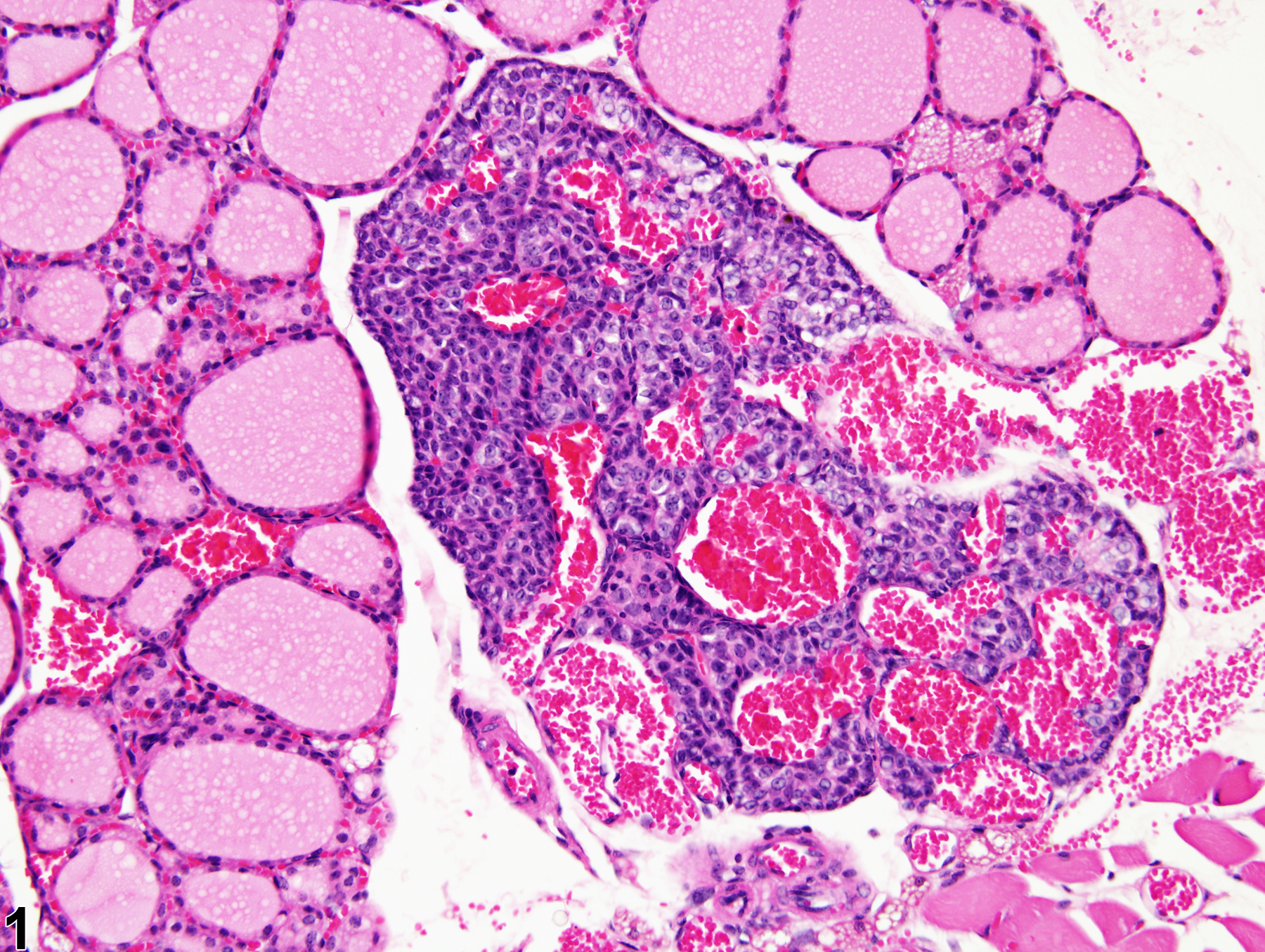
Parathyroid Gland - Angiectasis in a male BALB/c mouse from a subchronic study. Multiple dilated vascular structures filled with erythrocytes are present in the parathyroid.
All Images

Parathyroid Gland - Angiectasis in a male BALB/c mouse from a subchronic study. Multiple dilated vascular structures filled with erythrocytes are present in the parathyroid.
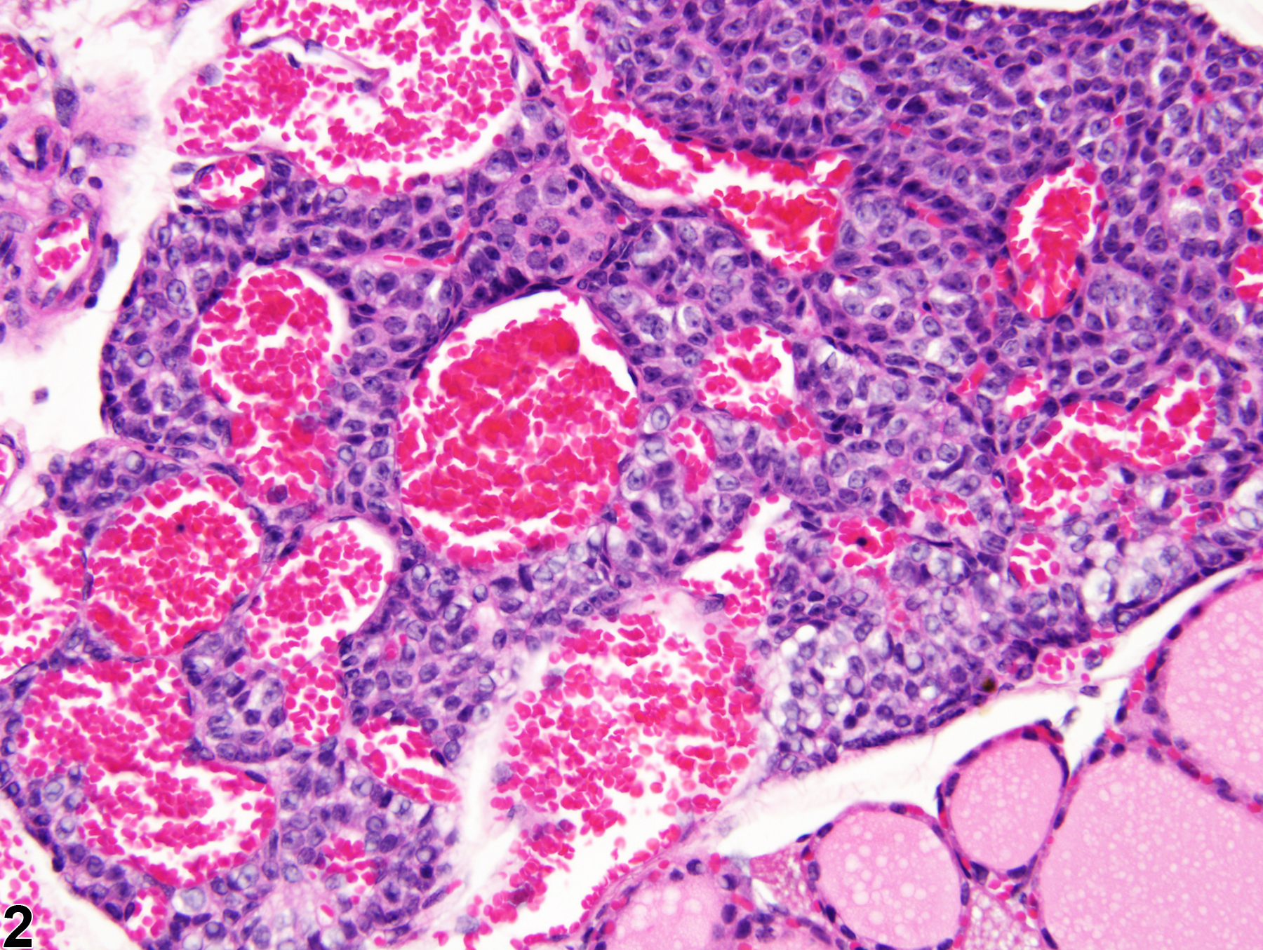
Parathyroid Gland - Angiectasis in a male BALB/c mouse from a subchronic study. This higher magnification of Figure 1 shows moderately and markedly dilated vascular spaces filled with erythrocytes.