U.S. flag

An official website of the United States government

Dot gov

The .gov means it's official.
Federal government websites often end in .gov or .mil. Before sharing sensitive information, make sure you're on a federal government site.

Https

The site is secure.
The https:// ensures that you are connecting to the official website and that any information you provide is encrypted and transmitted securely.

Skip to Main Navigation
Skip to Page Content
Skip to Atlas Navigation

Parathyroid Gland - Hyperplasia, Diffuse

Image of hyperplasia, diffuse in the parathyroid gland from a male F344/N rat in a chronic study
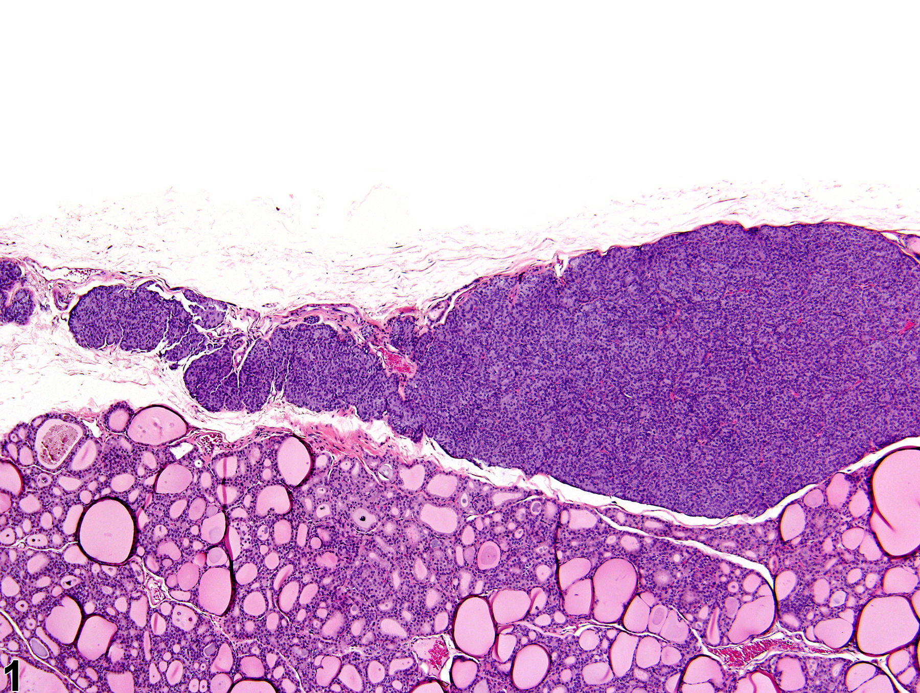
Parathyroid gland - Hyperplasia, Diffuse in a male F344/N rat from a chronic study. The parathyroid gland is increased in volume.
Figure 1 of 2
Image of hyperplasia, diffuse in the parathyroid gland from a male F344/N rat in a chronic study
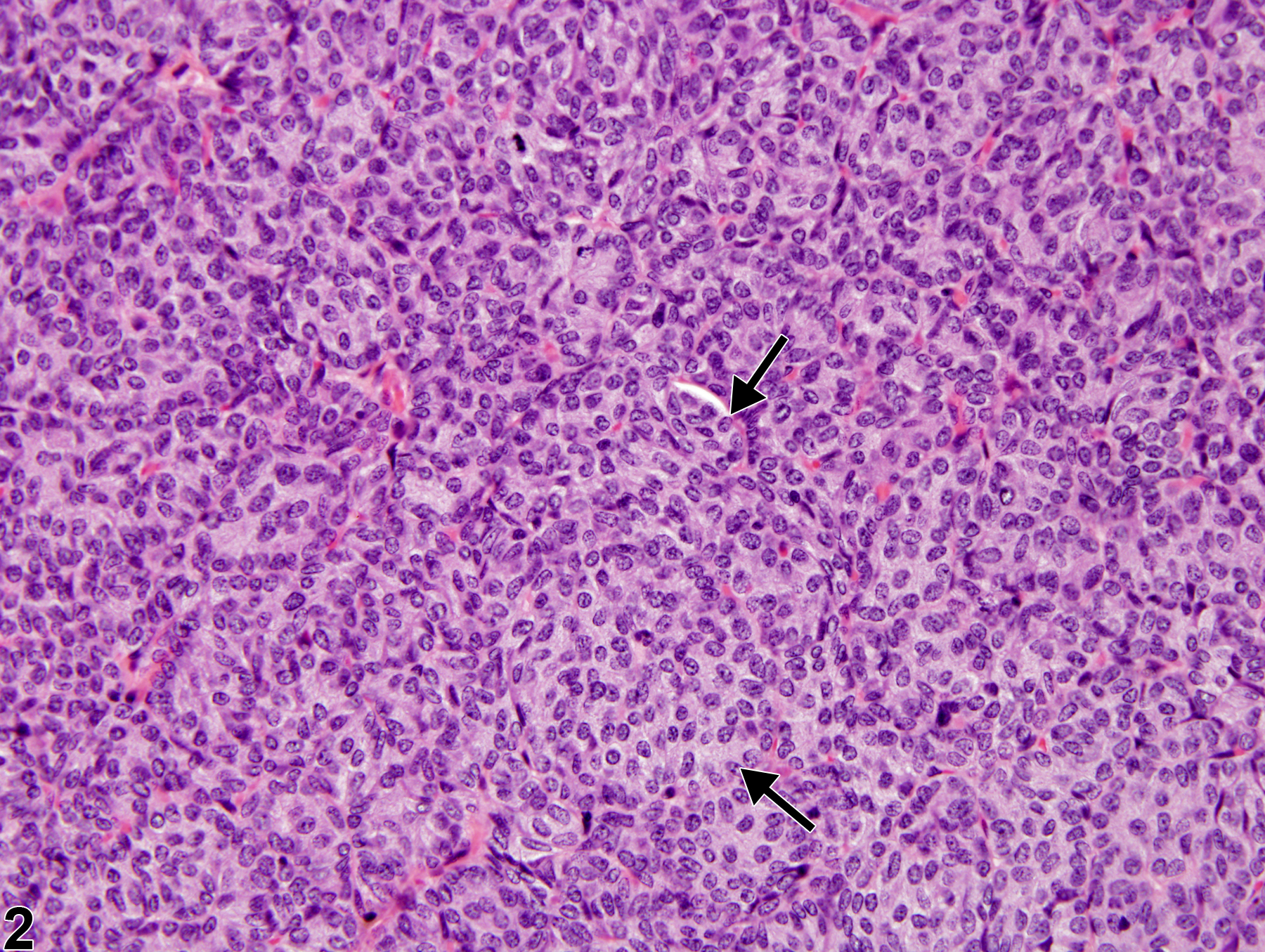
Parathyroid gland - Hyperplasia, Diffuse in a male F344/N rat from a chronic study. Enlarged nests of mildly hypertrophic parathyroid chief cells (arrows) have an increased cytoplasmic-to-nuclear ratio, and the intervening fibroreticular stroma is less apparent than normal.
Figure 2 of 2
next arrow

comment:

Parathyroid hyperplasia can be focal or diffuse and occurs in low incidence in rats and rarely in mice. Diffuse hyperplasia may be associated with renal failure and long-term dietary imbalance of calcium and/or phosphorus and is usually bilateral. Diffuse hyperplasia presents as a uniform increase in cellularity due to combined hypertrophy and hyperplasia. With diffuse hyperplasia, the normal cellular nests and cords are altered, with increased size of hyperplastic nests being most obvious. Severe hyperplasia will usually result in a grossly enlarged parathyroid but may be difficult to determine histologically if the tissue in the histologic sections is not well sampled. Distinction from parathyroid adenoma is based on the adenoma appearing as a discrete expansile lesion, usually unilateral, within the gland and with compression of adjacent parenchyma.

recommendation:

Hyperplasia should be diagnosed and given a severity grade whenever observed. If both parathyroids are involved, the diagnosis should be qualified as bilateral and the severity grade should be based on the more severely affected gland. If the pathologist feels the hyperplasia is associated with renal disease, it should be noted in the pathology narrative. Other lesions that may be associated with the hyperplasia, such as gastric or renal mineralization, should also be commented on in the pathology narrative.

references:

Capen CC, Rosol TJ. 1989. Recent advances in the structure and function of the parathyroid gland in animals and the effects of xenobiotics. Toxicol Pathol 17:333-345.
Abstract: http://www.ncbi.nlm.nih.gov/pubmed/2675284

Capen CC, DeLellis RA, Yarrington JT. 2002. Endocrine system. In: Handbook of Toxicologic Pathology, Vol 2 (Haschek WM, Rousseaux CG, Wallig MA, eds). Academic Press, New York, 681-783.
Abstract: http://www.sciencedirect.com/science/book/9780123302151

Rosol TJ, Capen CC. 1989. Tumors of the parathyroid gland and circulating parathyroid hormone-related protein associated with persistent hypercalcemia. Toxicol Pathol 17:346-356.
Full Text: http://tpx.sagepub.com/content/17/2/346.full.pdf

Seely JC, Hildebrandt PK.. 1990. Parathyroid gland. In: Pathology of the Fischer Rat: Reference and Atlas (Boorman GA, Eustis SL, Elwell MR, Montgomery CA, MacKenzie WF, eds). Academic Press, San Diego, 537-543.
Abstract: http://www.ncbi.nlm.nih.gov/nlmcatalog/9002563