

Endocrine System
Pituitary Gland - Cyst
Narrative
The majority of the literature dealing with pituitary cysts makes reference to a craniopharyngeal origin and further indicates that Rathke's pouch is part of the craniopharyngeal structure. Consequently, cysts of Rathke's pouch and craniopharyngeal cysts are synonymous. Determination of the origin of specific cysts may be difficult.
Capen CC. 1983. Functional and pathologic interrelationships of the pituitary gland and hypothalamus. In: Endocrine System (Jones TC, Mohr U, Hunt RD, eds). Springer, New York, 103-120.
Abstract: http://www.springer.com/medicine/pathology/book/978-3-642-96722-1Capen CC, DeLellis RA, Yarrington JT. 1991. Endocrine system. In: Handbook of Toxicologic Pathology (Haschek WM, Rousseaux CG, eds). Academic Press, New York, 697-705.
Abstract: http://www.sciencedirect.com/science/book/9780123302151Carlton WW, Gries CL. 1983. Cysts, pituitary; rat, mouse, and hamster. In: Endocrine System (Jones TC, Mohr U, Hunt RD, eds). Springer, New York, 161-163.
Abstract: http://www.springer.com/medicine/pathology/book/978-3-642-96722-1Gon G, Nakamura F, Ishikawa H. 1987. Cystlike structures derived from the marginal cells of Rathke's cleft in rat pituitary grafts. Cell Tissue Res 250:29-33.
Abstract: http://www.ncbi.nlm.nih.gov/pubmed/3652164Herrick E, McCormack S. 1952. The occurrence and nature of cysts in the pituitary of fowls and rats. Trans Kans Acad Sci 55:178-183.
Abstract: http://www.jstor.org/stable/3625875Iwata H, Hosoi M, Miyajima R, Yamamoto S, Mikami S, Yamakawa S, Enomoto M, Imazawa T, Mitsumori K. 2000. Morphogenesis of craniopharyngeal derivatives in the neurohypophysis of Fisher 344 rats: Abnormally developed epithelial tissues including parotid glands derived from the stomatodeum. Toxicol Pathol 28:568-574.
Abstract: http://www.ncbi.nlm.nih.gov/pubmed/10930044Kouki T, Imai H, Aoto K, Eto K, Shioda S, Kawamura K, Kikuyama S. 2001. Developmental origin of the rat adenohypophysis prior to the formation of Rathke's pouch. Development 128:959-963.
Abstract: http://www.ncbi.nlm.nih.gov/pubmed/11222149Lansdown AB, Grasso P 1971. Histological observations on a Rathke's cleft abnormality in a laboratory rat. J Comp Pathol 81:141-144.
Abstract: http://www.sciencedirect.com/science/article/pii/0021997571900661MacKenzie WF, Boorman GA. 1991. Pituitary gland. In: Pathology of the Fischer Rat: Reference and Atlas (Boorman G, Eustis S, Elwell M, Montgomery CA, MacKenzie W, eds). Academic Press, San Diego, 485-500.
Abstract: http://www.ncbi.nlm.nih.gov/nlmcatalog/9002563Mahler J, Elwell M. 1999. The pituitary gland. In: Pathology of the Mouse: Reference and Atlas (Maronpot RR, Boorman GA, Gaul BW, eds). Cache River Press, Vienna, IL, 491-508.
Morton D, Tekeli S. 1997. "Have you seen this?" Pituitary cysts in a mouse. Toxicol Pathol 25:333.
Full Text: http://tpx.sagepub.com/content/25/3/333.longQuintanar-Stephano A, Munoz Fernandez L, Quintanar JL, Kovacs K. 2001. Cysts in the rat adenohypophysis: Incidence and histology. Endocr Pathol 12:63-71.
Abstract: http://www.ncbi.nlm.nih.gov/pubmed/11478270Watanabe YG. 1991 The occurrence and developmental origin of epithelial cysts in the rat and mouse adenohypophysis. Arch Histol Cytol 54:511-518.
Abstract: http://www.ncbi.nlm.nih.gov/pubmed/1665339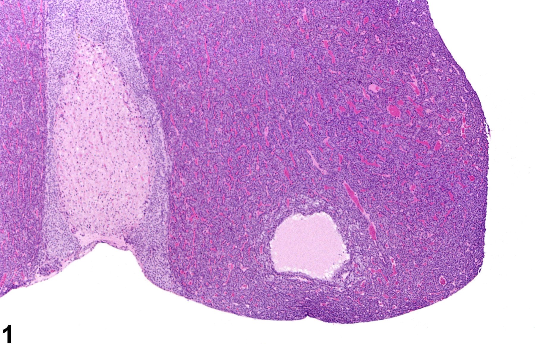
Pituitary Gland - Cyst in a female Harlan Sprague-Dawley rat from a chronic study. A cyst filled with pale eosinophilic proteinaceous fluid is present in the pars distalis.
All Images

Pituitary Gland - Cyst in a female Harlan Sprague-Dawley rat from a chronic study. A cyst filled with pale eosinophilic proteinaceous fluid is present in the pars distalis.
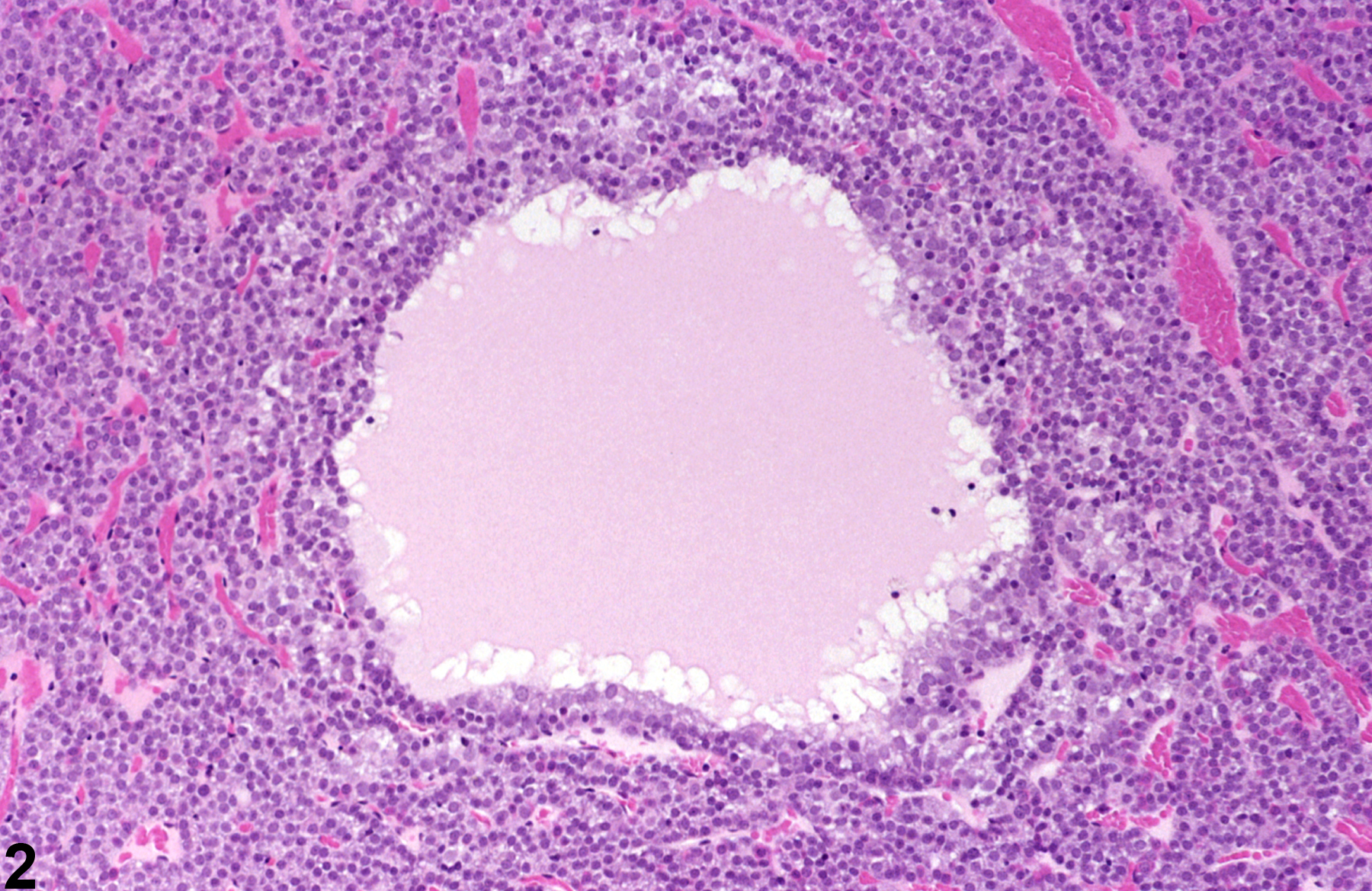
Pituitary Gland - Cyst in a female Harlan Sprague-Dawley rat from a chronic study. Higher magnification of Figure 1 shows that the cyst is partially lined by tall cuboidal ciliated epithelium.
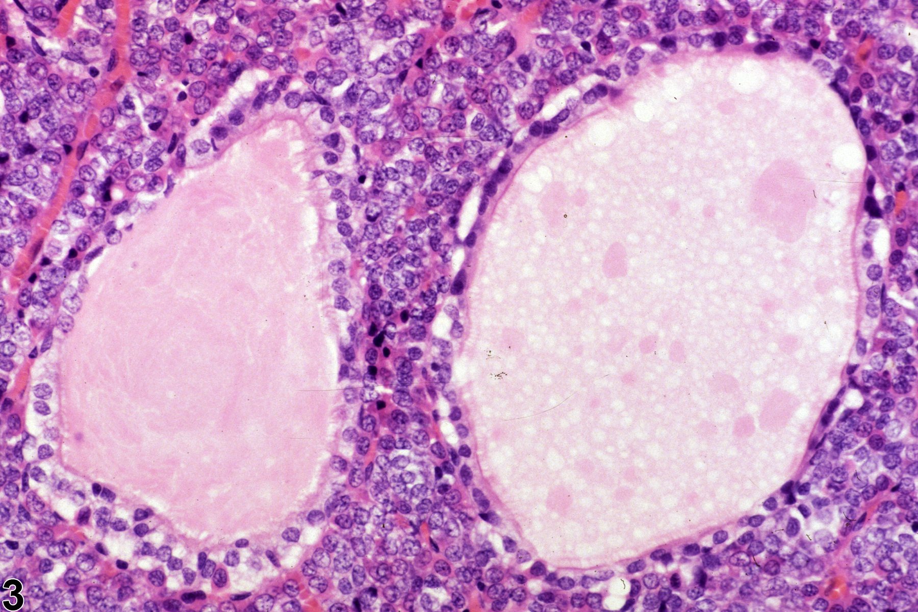
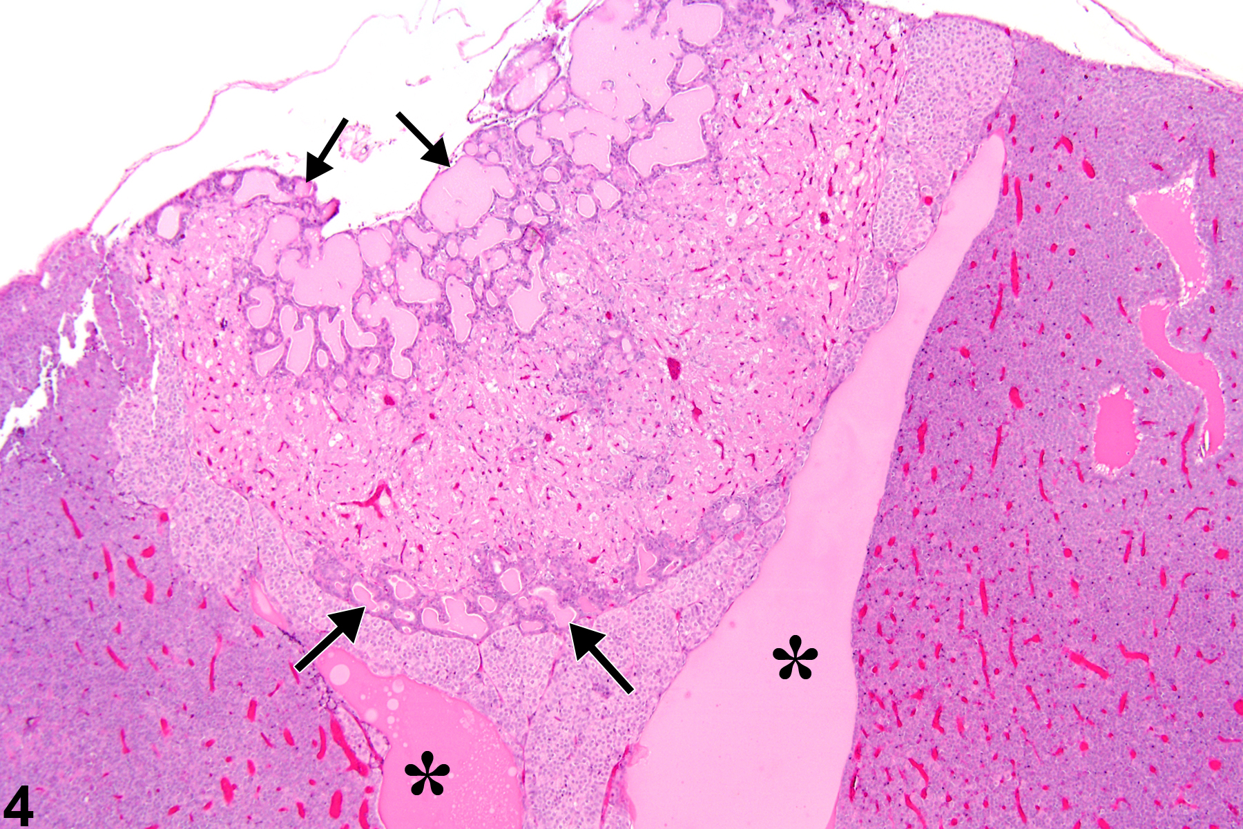
Pituitary Gland - Cyst in a male F344/n rat from a chronic study. The cysts in the pars distalis are lined by ciliated cuboidal cells and contain flocculant eosinophilic proteinaceous material.


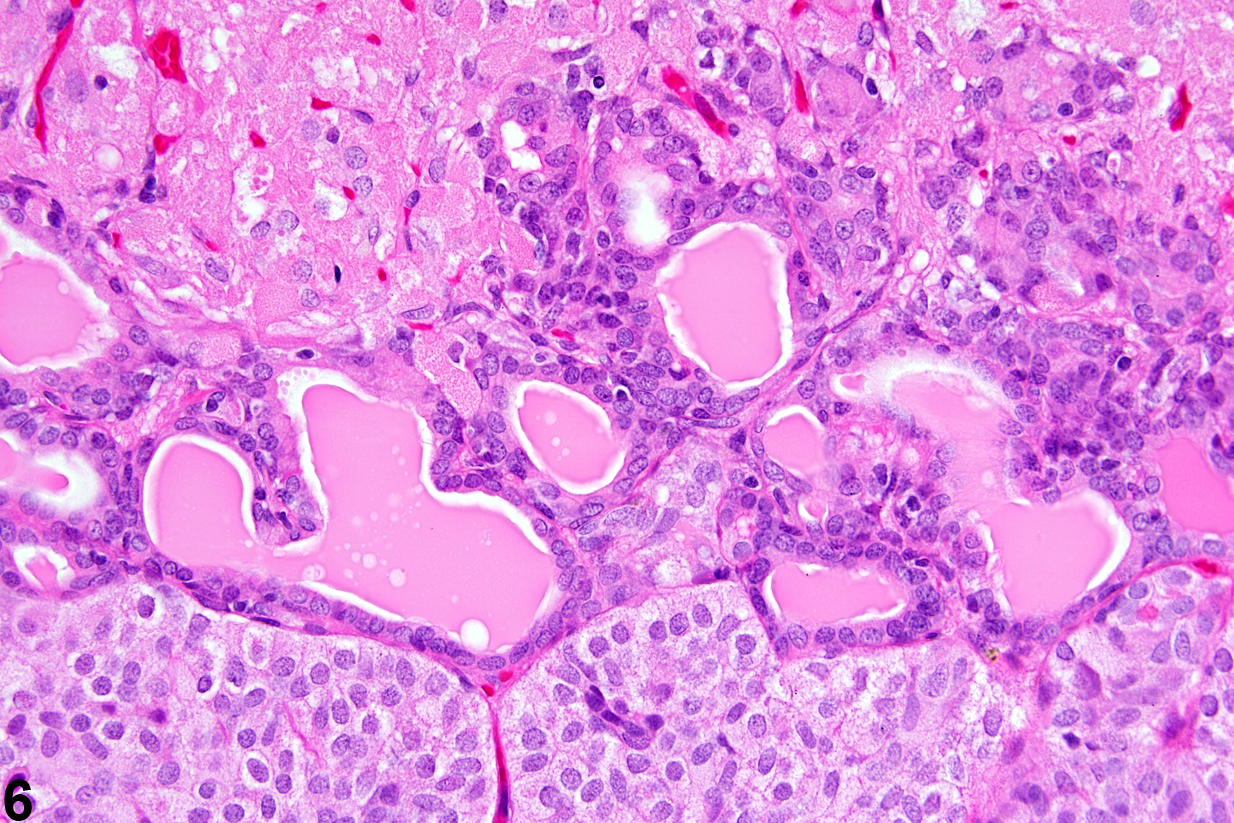
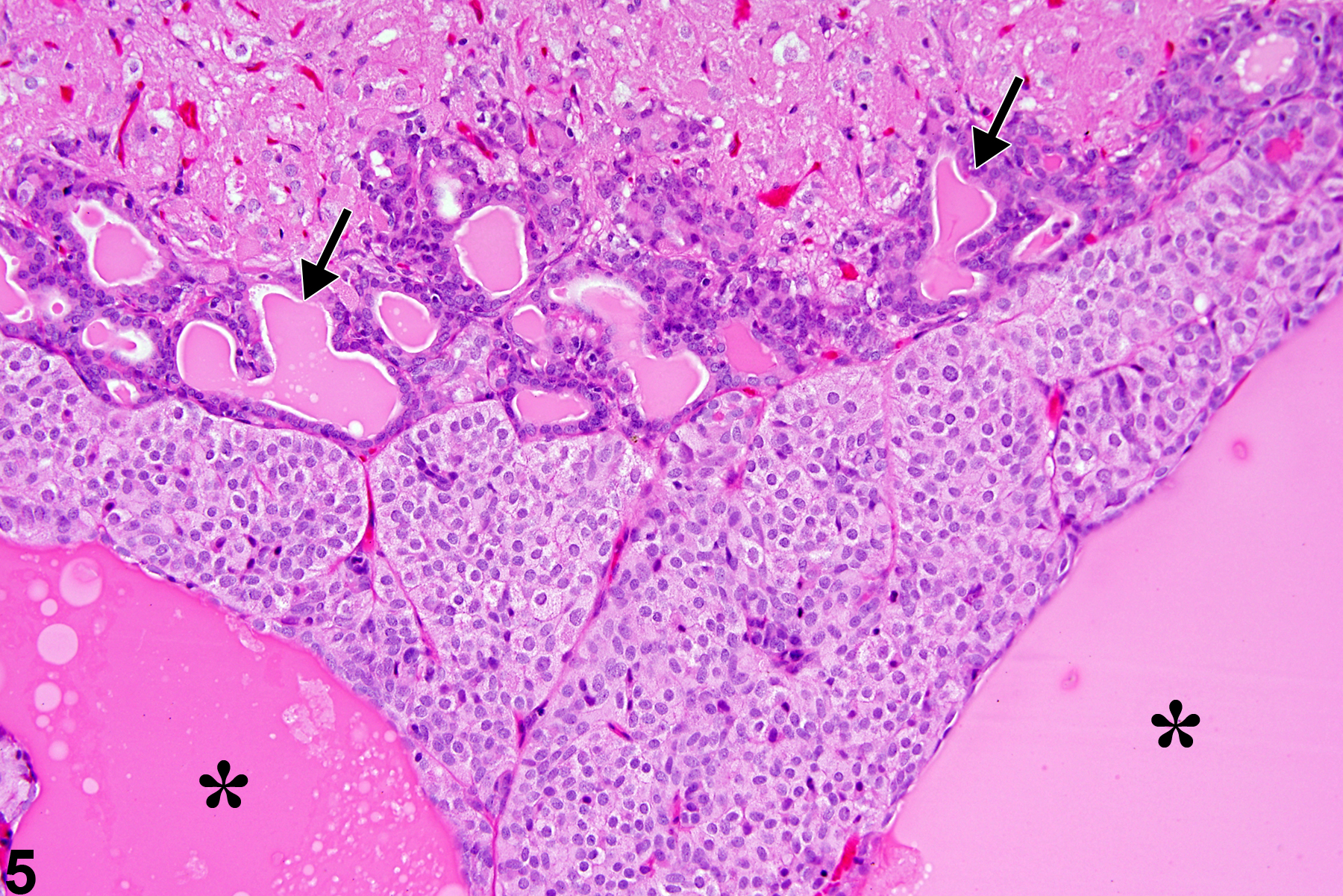
Pituitary Gland - Cysts, multiple in a female F344/N rat from a chronic study. Higher magnification of Figure 5 shows the epithelium-lined cysts are present in the pars nervosa.