

Endocrine System
Pituitary Gland, Pars Distalis - Atrophy
Narrative
Daniel PM, Prichard MM. 1956. Anterior pituitary necrosis; infarction of the pars distalis produced experimentally in the rat. Q J Exp Physiol Cogn Med Sci 41:215-229.
Full Text: http://ep.physoc.org/content/41/3/215.long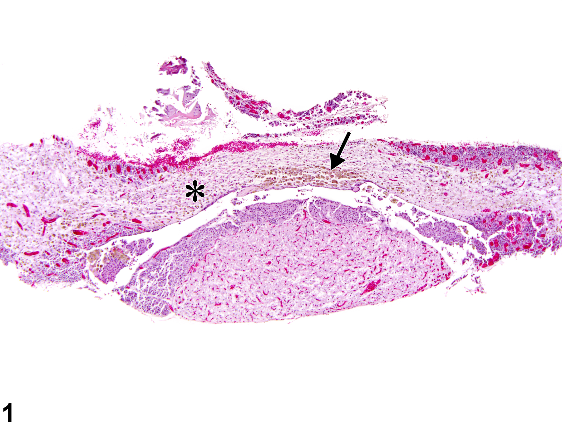
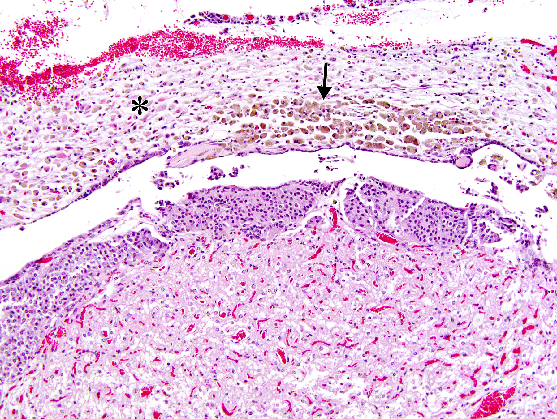
Pituitary Gland, Pars distalis - Atrophy in a female F344/N rat from a chronic study. The pars distalis (asterisk) is reduced in size and contains a focal aggregate of yellow-brown pigment (likely hemosiderin) within macrophages (arrow).
All Images

Pituitary Gland, Pars distalis - Atrophy in a female F344/N rat from a chronic study. The pars distalis (asterisk) is reduced in size and contains a focal aggregate of yellow-brown pigment (likely hemosiderin) within macrophages (arrow).

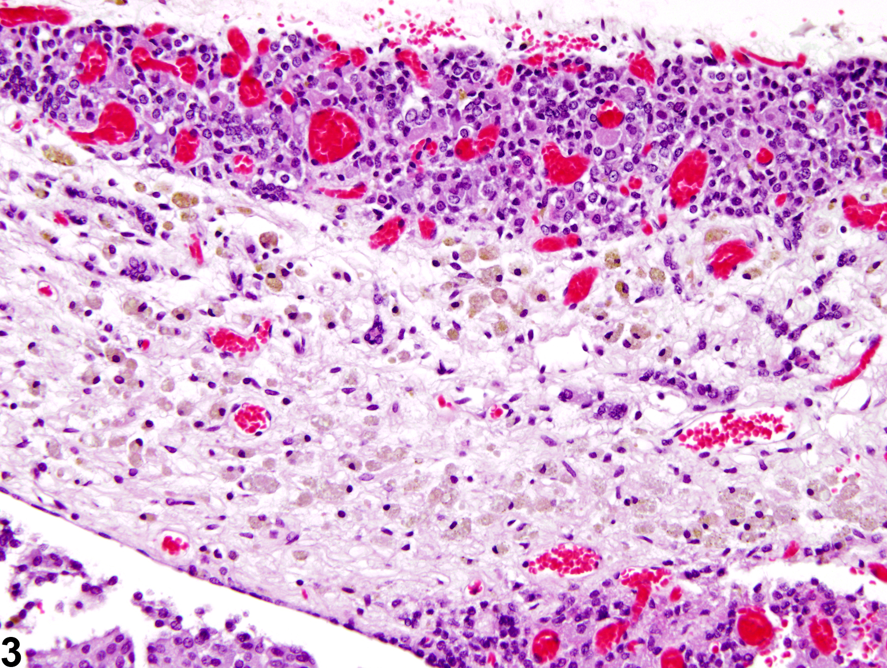
Pituitary Gland, Pars distalis - Atrophy in a female F344/N rat from a chronic study. Higher magnification of Figure 1 shows the reduction in the size of the pars distalis (asterisk) and the focal aggregate of macrophages (arrow) containing yellow-brown pigment (likely hemosiderin).

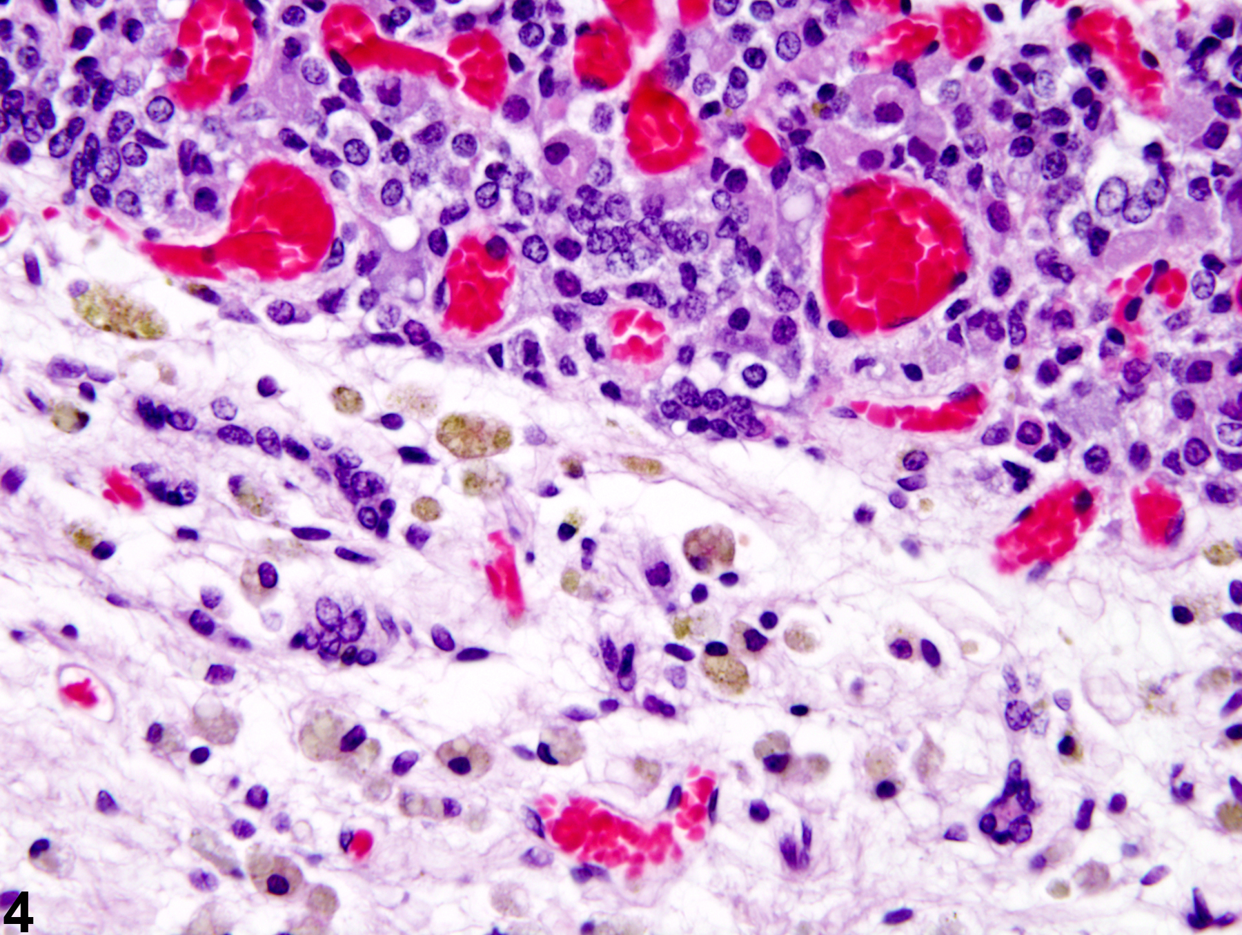
Pituitary Gland, Pars distalis - Atrophy in a female F344/N rat from a chronic study. Higher magnification of Figure 2 shows that the atrophic pars distalis has a paucity of cells and that macrophages containing yellow-brown pigment (likely hemosiderin) are present in the atrophic tissue.

Pituitary Gland, Pars distalis - Atrophy in a female F344/N rat from a chronic study. Higher magnification of Figure 3 shows the atrophic pars distalis with the pigment laden macrophages in greater detail.