

Endocrine System
Pituitary Gland, Rathke's Cleft - Dilation
Narrative
Capen CC. 1983. Functional and pathologic interrelationships of the pituitary gland and hypothalamus. In: Endocrine System (Jones TC, Mohr U, Hunt RD, eds). Springer, New York, 103-120.
Abstract: http://www.springer.com/medicine/pathology/book/978-3-642-96722-1Capen CC, DeLellis RA, Yarrington JT. 1991. Endocrine system. In: Handbook of Toxicologic Pathology (Haschek WM, Rousseaux CG, eds). Academic Press, New York, 697-705.
Abstract: http://www.sciencedirect.com/science/book/9780123302151Carlton WW, Gries CL. 1983. Cysts, pituitary; rat, mouse, and hamster. In: Endocrine System (Jones TC, Mohr U, Hunt RD, eds). Springer, New York, 161-163.
Abstract: http://www.springer.com/medicine/pathology/book/978-3-642-96722-1Gon G, Nakamura F, Ishikawa H. 1987. Cystlike structures derived from the marginal cells of Rathke's cleft in rat pituitary grafts. Cell Tissue Res 250:29-33.
Abstract: http://www.ncbi.nlm.nih.gov/pubmed/3652164Herrick E, McCormack S. 1952. The occurrence and nature of cysts in the pituitary of fowls and rats. Trans Kans Acad Sci 55:178-183.
Abstract: http://www.jstor.org/stable/3625875Iwata H, Hosoi M, Miyajima R, Yamamoto S, Mikami S, Yamakawa S, Enomoto M, Imazawa T, Mitsumori K. 2000. Morphogenesis of craniopharyngeal derivatives in the neurohypophysis of Fisher 344 rats: Abnormally developed epithelial tissues including parotid glands derived from the stomatodeum. Toxicol Pathol 28:568-574.
Abstract: http://www.ncbi.nlm.nih.gov/pubmed/10930044Kouki T, Imai H, Aoto K, Eto K, Shioda S, Kawamura K, Kikuyama S. 2001. Developmental origin of the rat adenohypophysis prior to the formation of Rathke's pouch. Development 128:959-963.
Abstract: http://www.ncbi.nlm.nih.gov/pubmed/11222149Lansdown AB, Grasso P 1971. Histological observations on a Rathke's cleft abnormality in a laboratory rat. J Comp Pathol 81:141-144.
Abstract: http://www.sciencedirect.com/science/article/pii/0021997571900661MacKenzie WF, Boorman GA. 1991. Pituitary gland. In: Pathology of the Fischer Rat: Reference and Atlas (Boorman G, Eustis S, Elwell M, Montgomery CA, MacKenzie W, eds). Academic Press, San Diego, 485-500.
Abstract: http://www.ncbi.nlm.nih.gov/nlmcatalog/9002563Morton D, Tekeli S. 1997. "Have you seen this?" Pituitary cysts in a mouse. Toxicol Pathol 25:333.
Full Text: http://tpx.sagepub.com/content/25/3/333.longQuintanar-Stephano A, Munoz Fernandez L, Quintanar JL, Kovacs K. 2001. Cysts in the rat adenohypophysis: Incidence and histology. Endocr Pathol 12:63-71.
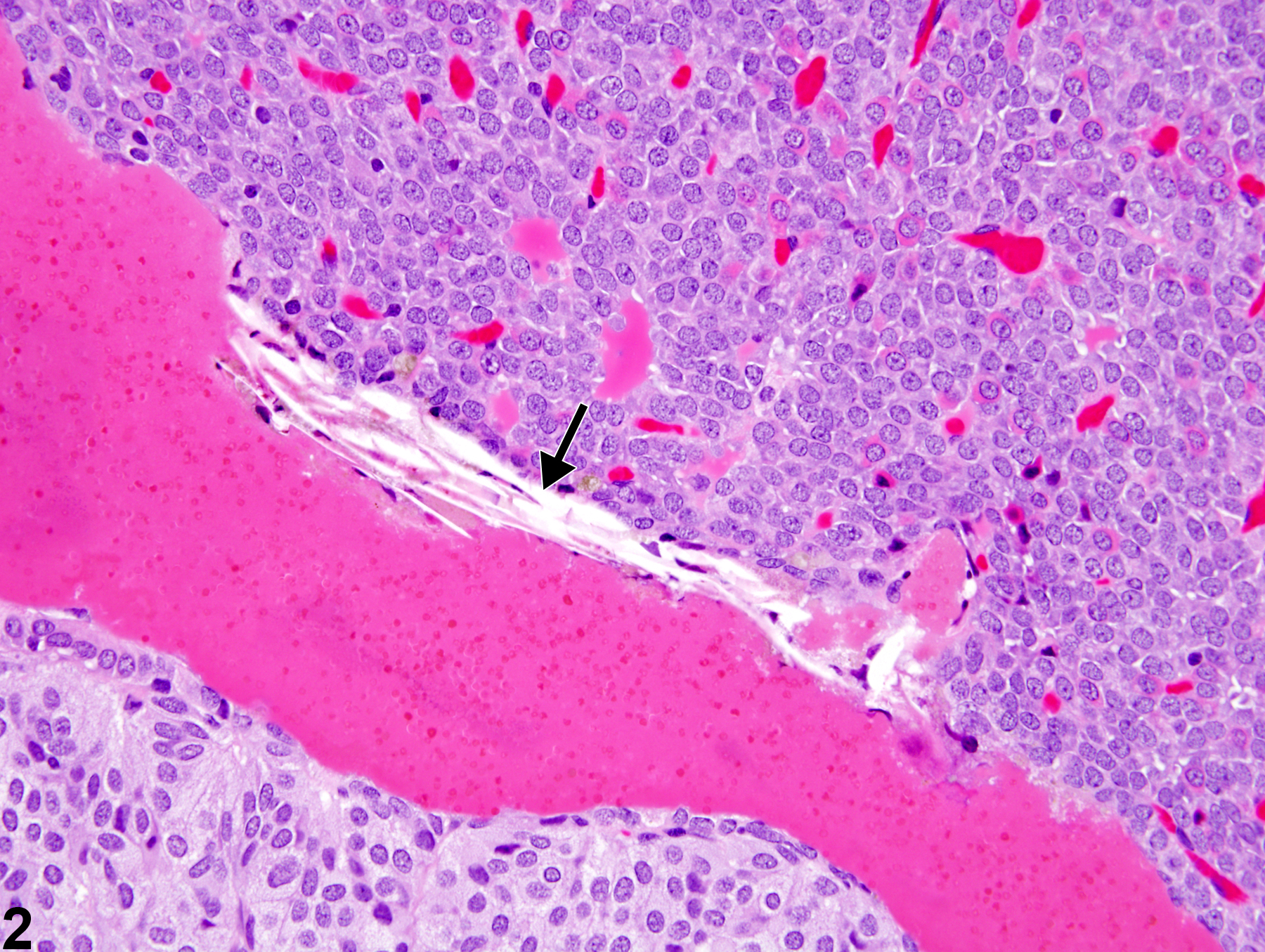
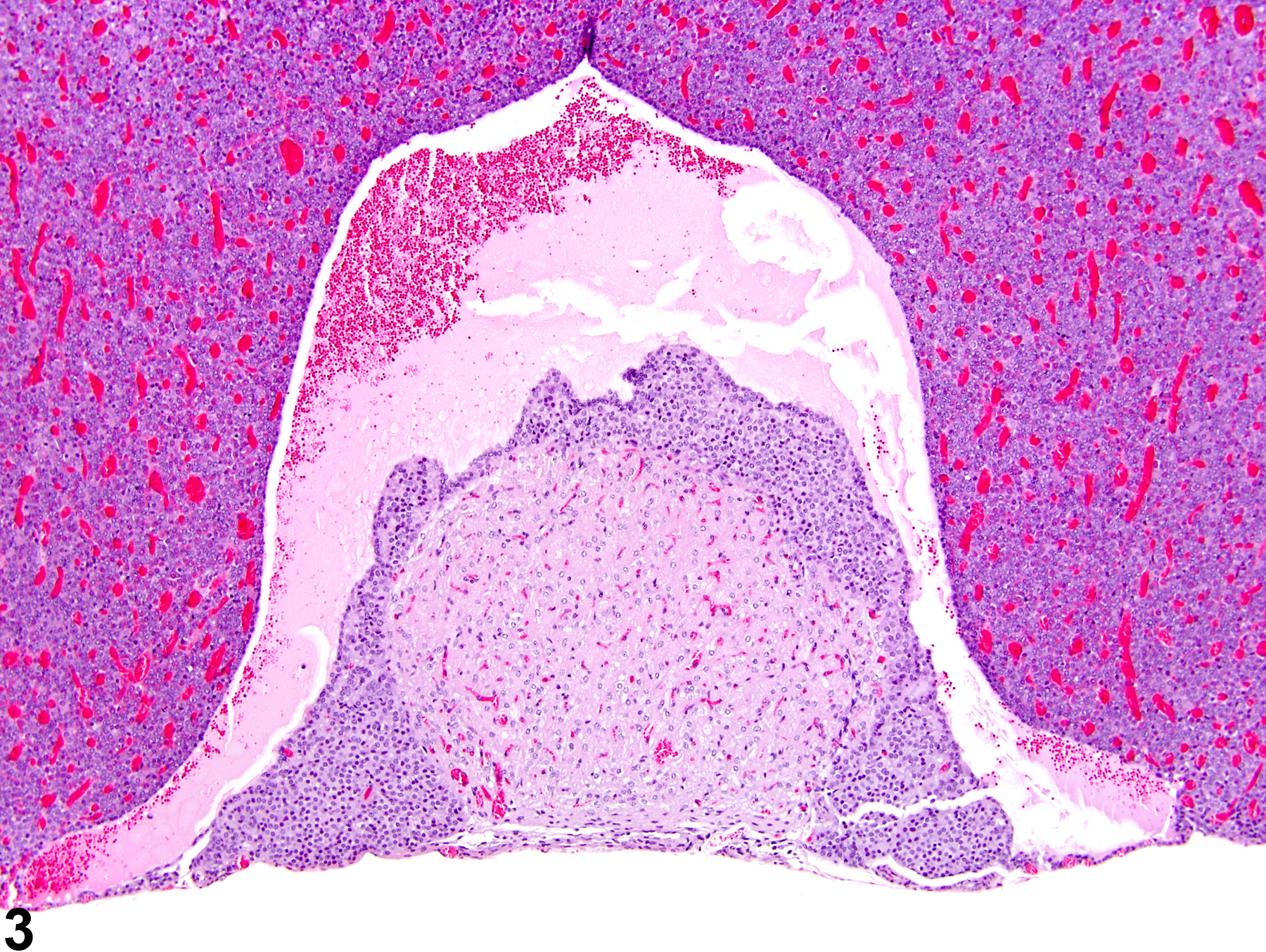
Abstract: http://www.ncbi.nlm.nih.gov/pubmed/11478270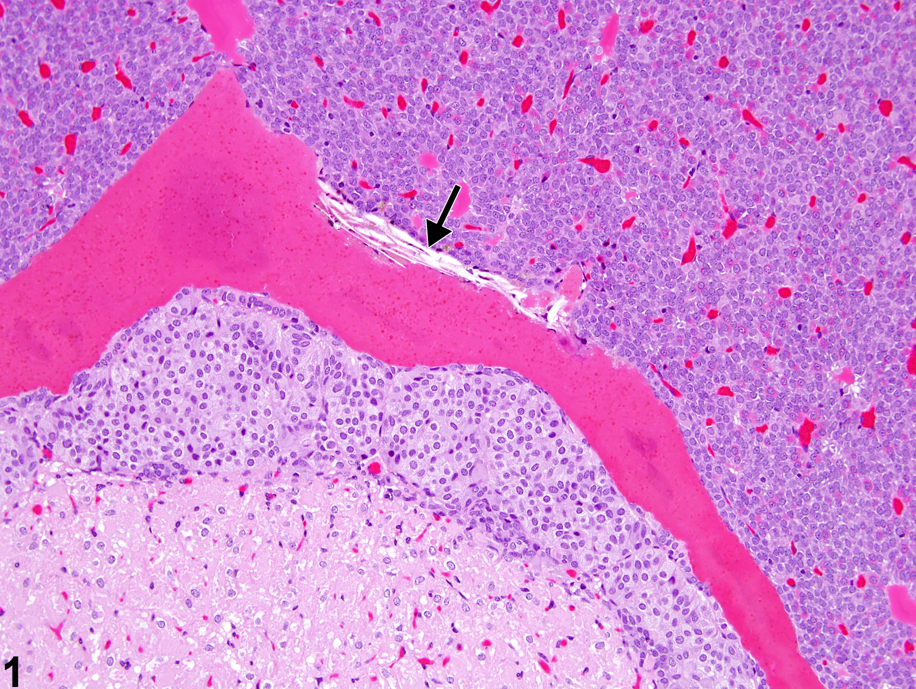 Pituitary, Rathke's cleft - Dilation in a female F344/N rat from a chronic study. The dilated Rathke's cleft is filled with intensely eosinophilic proteinaceous material with cholesterol clefts (arrow) present at the edge of the dilated cleft.
All Images


