

Endocrine System
Thyroid Gland, Follicular Cell - Hypertrophy
Narrative
Capen CC, Martin SL. 1989. The effects of xenobiotics on the structure and function of thyroid follicular and C-cells. Toxicol Pathol 17:266-293.
Abstract: http://www.ncbi.nlm.nih.gov/pubmed/2675279Capen CC, DeLellis RA, Yarrington JT. 2002. Endocrine system. In: Handbook of Toxicologic Pathology, Vol 2 (Haschek WM, Rousseaux CG, Wallig MA, eds). Academic Press, New York, 681–783.
Abstract: http://www.sciencedirect.com/science/book/9780123302151Collins WT, Capen CC, Kasza L, Carter C, Dailey RE. 1977. Effect of polychlorinated biphenyl (PCB) on the thyroid gland of rats. Ultrastructural and biochemical investigations. Am J Pathol 89:119-136.
Abstract: http://www.ncbi.nlm.nih.gov/pubmed/199066Greaves P. 2007. Histopathology of Preclinical Toxicity Studies: Interpretation and Relevance in Drug Safety Evaluation, 3rd ed. Academic Press, Amsterdam, 819-839.
Abstract: http://www.sciencedirect.com/science/book/9780444527714Hardisty JF, Boorman GA. 1990. Thyroid gland. In: Pathology of the Fischer Rat: Reference and Atlas (Boorman GA, Eustis SL, Elwell MR, Montgomery CA, MacKenzie WF, eds). Academic Press, San Diego, 519–536.
Abstract: http://www.ncbi.nlm.nih.gov/nlmcatalog/9002563Hood A, Hashmi R, Klaassen CD. 1999. Effects of microsomal enzyme inducers on thyroid-follicular cell proliferation, hyperplasia, and hypertrophy. Toxicol Appl Pharmacol 160:163-170.
Abstract: http://www.ncbi.nlm.nih.gov/pubmed/10527915Stevens JT. 1985. Effect of chemicals on the thyroid gland. In: Endocrine Toxicology (Thomas JA, Korach KS, McLachlan JA, eds). Raven Press, New York, 135–147.
Abstract: http://informahealthcare.com/doi/book/10.3109/9781420093100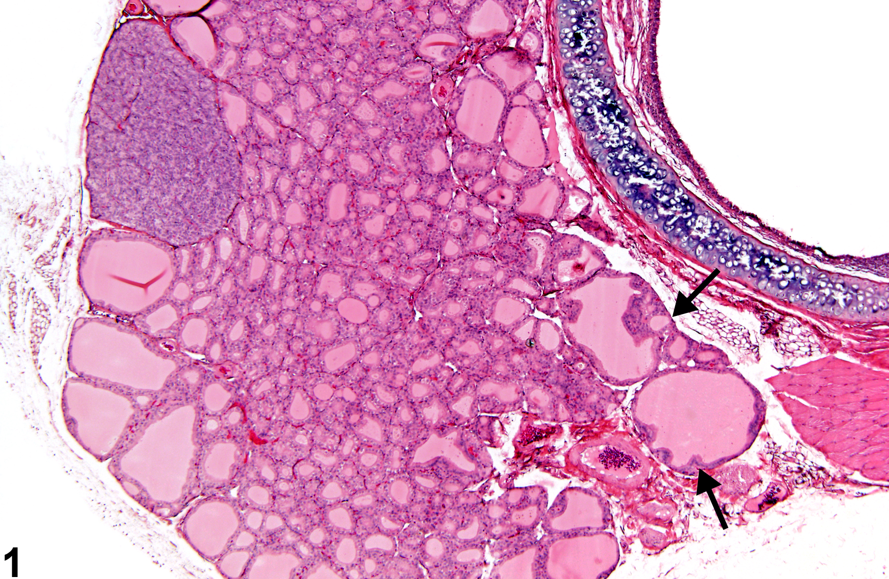
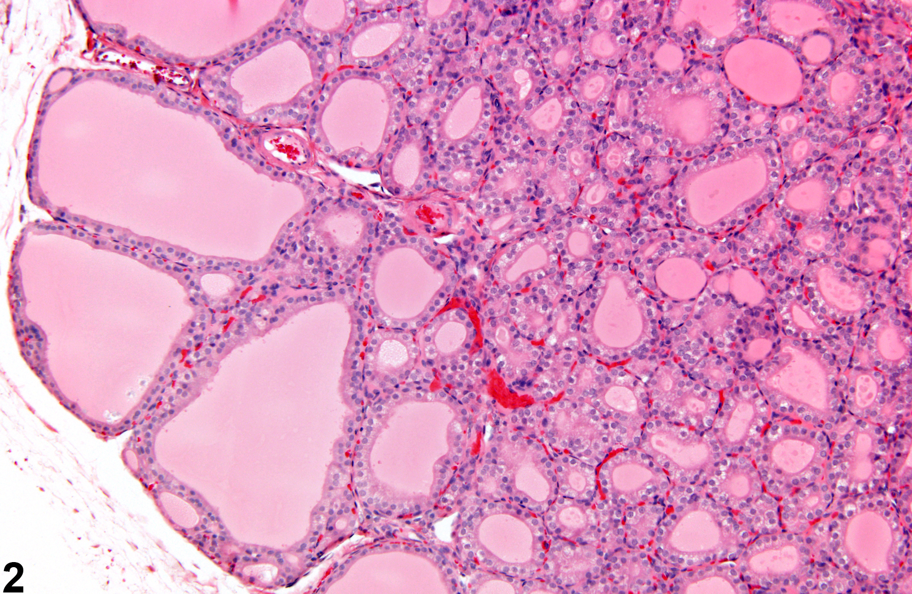
Thyroid Gland, Follicular Cell - Hypertrophy in a male F344/N rat from a subchronic study. Focal hyperplasia is present in two adjacent follicles (arrows).
All Images

Thyroid Gland, Follicular Cell - Hypertrophy in a male F344/N rat from a subchronic study. Focal hyperplasia is present in two adjacent follicles (arrows).

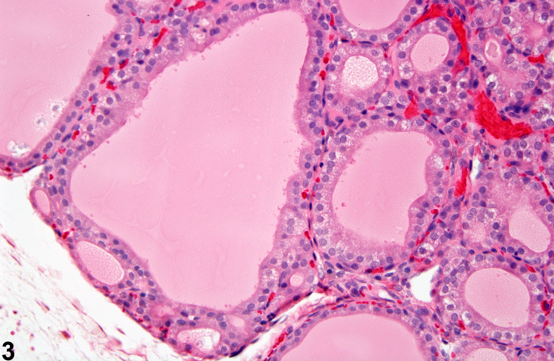
Thyroid Gland, Follicular Cell - Hypertrophy in a male F344/N rat from a subchronic study. Higher magnification of Figure 1 shows the epithelium lining the follicles is cuboidal to columnar.

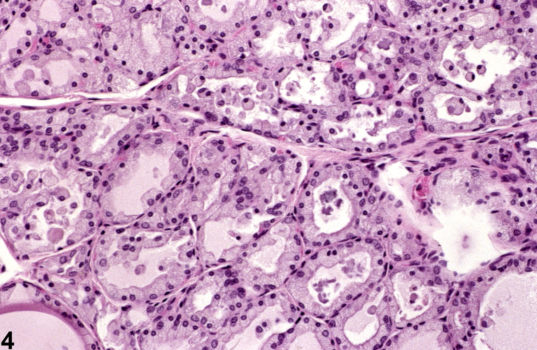
Thyroid Gland, Follicular Cell - Hypertrophy in a male F344/N rat from a subchronic study. The cuboidal to columnar lining epithelium contains small, clear, cytoplasmic vacuoles.

Thyroid Gland, Follicular Cell - Hypertrophy in a female Sprague-Dawley rat from a chronic study. Follicles are smaller than normal, are lined by cuboidal epithelium, and contain amphophilic, flocculant-appearing material and exfoliated epithelial cells.