

Endocrine System
Thyroid Gland, Follicle - Pigment
Narrative
Greaves P. 2007. Histopathology of Preclinical Toxicity Studies: Interpretation and Relevance in Drug Safety Evaluation, 3rd ed. Academic Press, Amsterdam, 819-839.
Abstract: http://www.sciencedirect.com/science/book/9780444527714Hardisty JF, Boorman GA. 1990. Thyroid gland. In: Pathology of the Fischer Rat: Reference and Atlas (Boorman GA, Eustis SL, Elwell MR, Montgomery CA, MacKenzie WF, eds). Academic Press, San Diego, 519–536.
Abstract: http://www.ncbi.nlm.nih.gov/nlmcatalog/9002563Hardisty JF, Boorman GA. 1999. Thyroid and parathyroid glands. In: Pathology of the Mouse: Reference and Atlas (Maronpot RR, Boorman GA, Gaul BW, eds). Cache River Press, Vienna, IL, 537–554.
Tajima K, Miyagawa J, Nakajima H, Shimizu M, Katayama S, Mashita K, Tarui S. 1985. Morphological and biochemical studies on minocycline-induced black thyroid in rats. Toxicol Appl Pharmacol 81:393-400.
Abstract: http://www.ncbi.nlm.nih.gov/pubmed/3001973Ward JM, Stinson SF, Hardisty JF, Cockrell BY, Hayden DW. 1979. Neoplasms and pigmentation of thyroid glands in F344 rats exposed to 2,4-diaminoanisole sulfate, a hair dye component. J Natl Cancer Inst 62:1067-1073.
Abstract: http://www.ncbi.nlm.nih.gov/pubmed/285280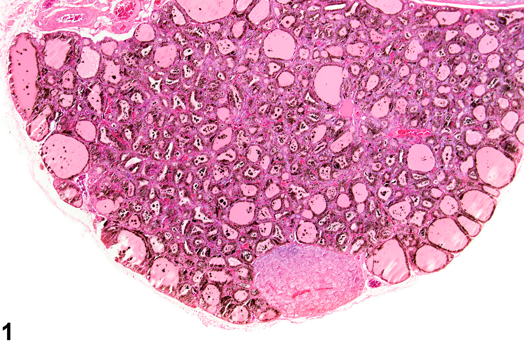
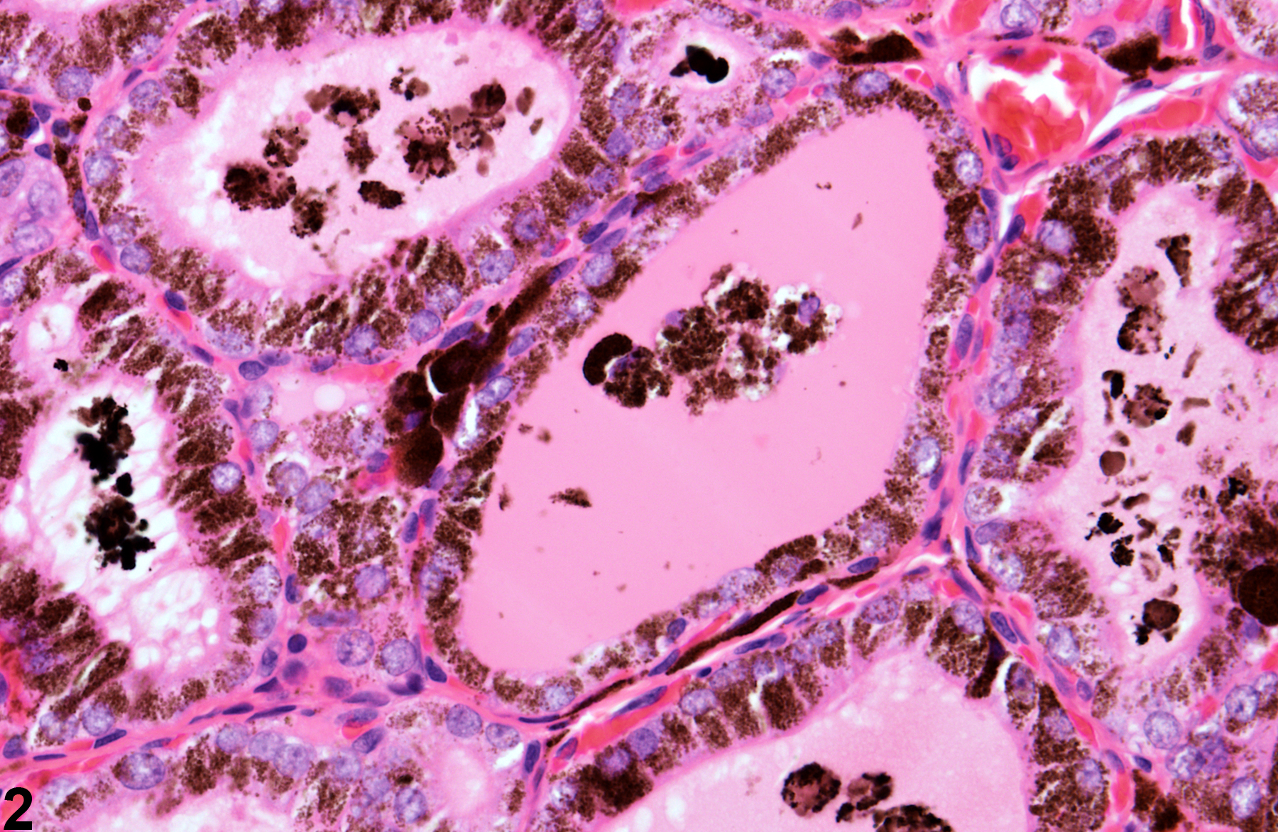
Thyroid gland, Follicle - Pigment in a male F344/N rat from a subchronic study. Dense pigment deposits are present in the follicular epithelium and colloid.
All Images

Thyroid gland, Follicle - Pigment in a male F344/N rat from a subchronic study. Dense pigment deposits are present in the follicular epithelium and colloid.

Thyroid gland, Follicle - Pigment in a male F344/N rat from a subchronic study. Higher magnification of the pigment deposits in the follicular epithelium colloid in Figure 1.

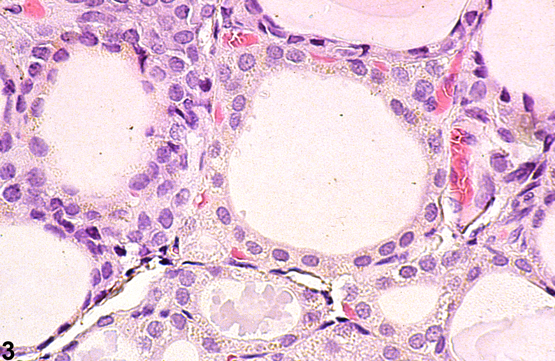
Thyroid gland, Follicle - Pigment in a male F344/N rat from a chronic study. Fine, brown, granular pigment is present within the follicular epithelium.

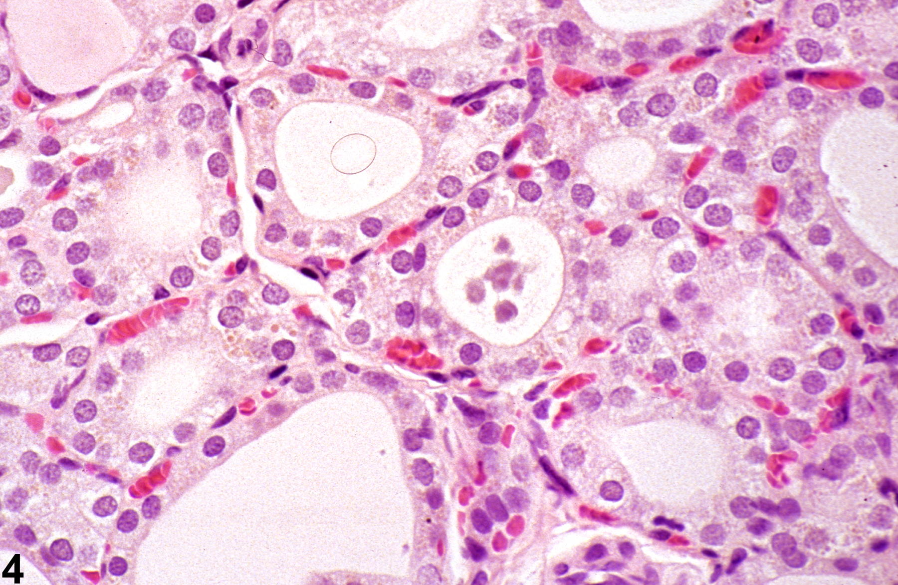
Thyroid gland, Follicle - Pigment in a male F344/N rat from a chronic study. Fine, granular, brown pigment is present within the follicular epithelium.