U.S. flag

An official website of the United States government

Dot gov

The .gov means it's official.
Federal government websites often end in .gov or .mil. Before sharing sensitive information, make sure you're on a federal government site.

Https

The site is secure.
The https:// ensures that you are connecting to the official website and that any information you provide is encrypted and transmitted securely.

Skip to Main Navigation
Skip to Page Content
Skip to Atlas Navigation

Thyroid Gland - Inflammation

Image of inflammation in the thyroid gland from a female Tg.AC (FVB/N) mouse in a chronic study
Thyroid Gland - Inflammation, chronic in a female Tg.AC (FVB/N) mouse from a subchronic study. Small aggregates of lymphoid cells are present at multiple sites between follicles.
Figure 1 of 3
Image of inflammation in the thyroid gland from a female Tg.AC (FVB/N) mouse in a chronic study
Thyroid Gland - Inflammation, chronic in a female Tg.AC (FVB/N) mouse from a subchronic study. Higher magnification of the small aggregates of lymphoid cells in Figure 1.
Figure 2 of 3
Image of inflammation in the thyroid gland from a female Tg.AC (FVB/N) mouse in a chronic study
Thyroid Gland - Inflammation, chronic in a female Tg.AC (FVB/N) mouse from a subchronic study. A perivascular accumulation of lymphoid cells is present.
Figure 3 of 3
next arrow

comment:

NTP studies have five standard categories of inflammation: acute, suppurative, chronic, chronic-active, and granulomatous. In acute inflammation, the predominant infiltrating cell is the neutrophil, though fewer macrophages and lymphocytes may also be present. There may also be evidence of edema or hyperemia. The neutrophil is also the predominant infiltrating cell type in suppurative inflammation, but they are aggregated, and many of them are degenerate (suppurative exudate). Cell debris from both the resident cell populations and infiltrating leukocytes, proteinaceous fluid containing fibrin, fewer macrophages, occasional lymphocytes or plasma cells, and, possibly, an infectious agent may also be present in within the exudate. Grossly, these lesions would be characterized by the presence of pus. The tissue surrounding the exudate may have fibroblasts, fibrous connective tissue, and mixed inflammatory cells, depending on the chronicity of the lesion. Lymphocytes predominate in chronic inflammation. Lymphocytes also predominate in chronic-active inflammation, but there are also a significant number of neutrophils. Both lesions may contain macrophages. Granulomatous inflammation is another form of chronic inflammation, but this diagnosis requires the presence of a significant number of aggregated, large, activated macrophages, epithelioid macrophages, or multinucleated giant cells.

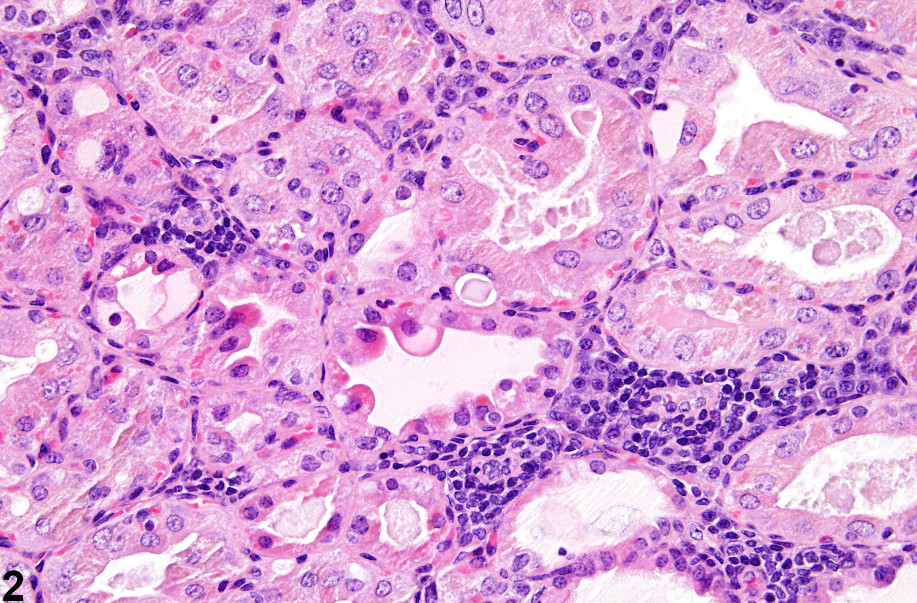
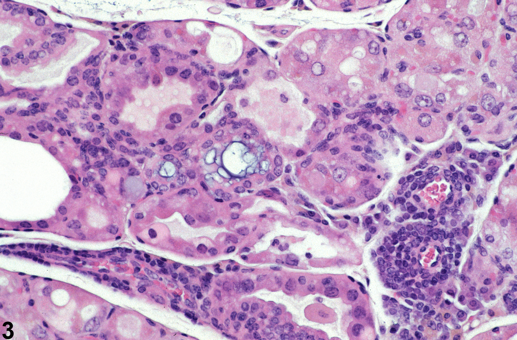
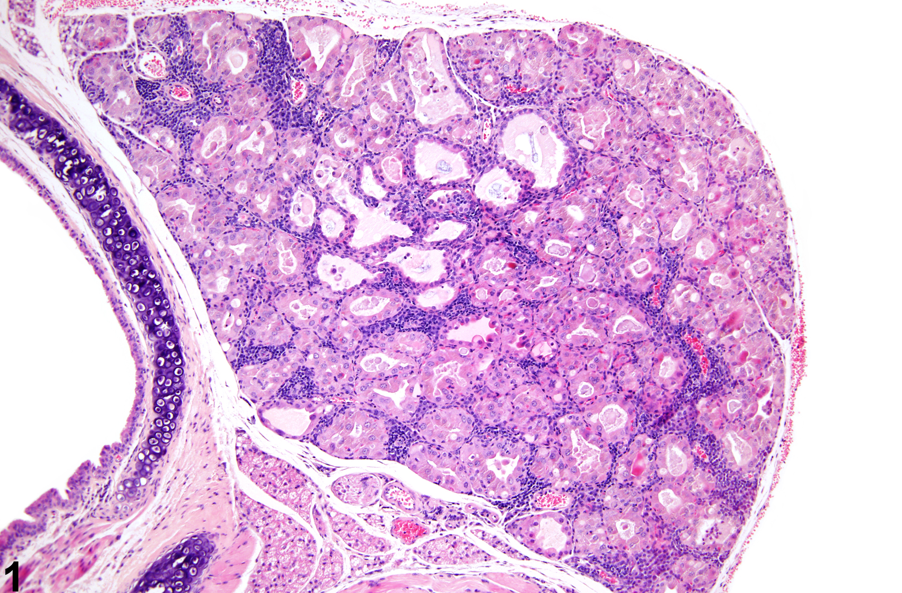
Spontaneously occurring cellular infiltrates are rare in thyroid glands of mice and rats. Multiple changes are present in the thyroids of these treated mice ( Figure 1image opens in a pop-up window , Figure 2image opens in a pop-up window , and Figure 3image opens in a pop-up window ). Lymphoid cell infiltrates are present between follicles ( Figure 1image opens in a pop-up window and Figure 2image opens in a pop-up window ) and in perivascular spaces ( Figure 3image opens in a pop-up window ). Additionally there is hypertrophy, degeneration, and necrosis of follicular epithelium.

recommendation:

Inflammation of the thyroid gland should be diagnosed and given a severity grade whenever present. The type of inflammation should be indicated in the diagnosis with a modifier (e.g., thyroid gland - Inflammation, chronic). In the absence of other changes characteristic of inflammation (e.g., edema, necrosis, degeneration), a diagnosis of cellular infiltrate with the cell type identified (e.g., thyroid gland - infiltrate, lymphoid) may be preferred. Other changes (e.g., colloid depletion, follicular cell degeneration, necrosis) not be diagnosed separately unless warranted by their severity. These other changes can be described in the pathology narrative. The diagnosis should indicate whether the inflammation is bilateral, with the severity grade based on the more severely affected thyroid.

references:

Greaves P. 2007. Histopathology of Preclinical Toxicity Studies: Interpretation and Relevance in Drug Safety Evaluation, 3rd ed. Academic Press, Amsterdam, 819-839.
Abstract: http://www.sciencedirect.com/science/book/9780444527714

Hardisty JF, Boorman GA. 1999. Thyroid and parathyroid glands. In: Pathology of the Mouse: Reference and Atlas (Maronpot RR, Boorman GA, Gaul BW, eds). Cache River Press, Vienna, IL, 537–554.
Abstract: http://www.cacheriverpress.com/books/pathmouse.htm